. 2007 Oct 18:7:196.
doi: 10.1186/1471-2148-7-196.

Species-specific evolution of immune receptor tyrosine based activation motif-containing CEACAM1-related immune receptors in the dog

Affiliations

Species-specific evolution of immune receptor tyrosine based activation motif-containing CEACAM1-related immune receptors in the dog

Robert Kammerer et al. BMC Evol Biol. .

Abstract

Background: Although the impact of pathogens on the evolution of the mammalian immune system is still under debate, proteins, which both regulate immune responses and serve as cellular receptors for pathogens should be at the forefront of pathogen-driven host evolution. The CEA (carcinoembryonic antigen) gene family codes for such proteins and indeed shows tremendous species-specific variation between human and rodents. Since little is known about the CEA gene family in other lineages of placental mammals, we expected to gain new insights into the evolution of the rapidly diverging CEA family by analyzing the CEA family of the dog.

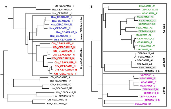
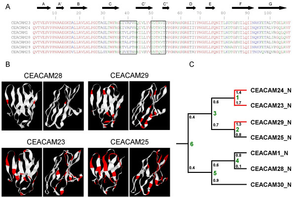
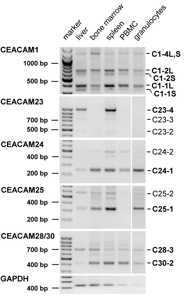
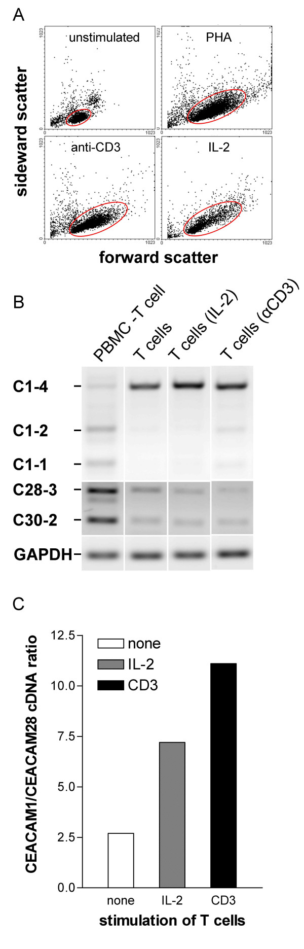
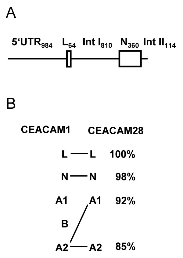
Results: Here we describe the complete CEA gene family in the dog. We found that the gene coding for the ITIM-bearing immunoregulatory molecule CEACAM1 gave rise to a recent expansion of the canine CEA gene family by gene duplication, similar to that previously found in humans and mice. However, while the murine and human CEACAMs (carcinoembryonic antigen-related cell adhesion molecules) are predominantly secreted and GPI-anchored, respectively, in the dog, most of the CEACAMs represent ITAM-bearing transmembrane proteins. One of these proteins, CEACAM28, exhibits nearly complete sequence identity with the ligand-binding N domain of CEACAM1, but antagonizing signaling motifs in the cytoplasmic tail. Comparison of nonsynonymous and synonymous substitutions indicates that the CEACAM28 N domain is under the strongest purifying selection of all canine CEACAM1-related CEACAMs. In addition, CEACAM28 shows a similar expression pattern in resting immune cells and tissues as CEACAM1. However, upon activation CEACAM28 mRNA and CEACAM1 mRNA are differentially regulated.

Conclusion: Thus, CEACAM1 and CEACAM28 are the first paired immune receptors identified within the CEA gene family, which are expressed on T cells and are most likely involved in the fine-tuning of T cell responses. The direction of gene conversion accompanied by purifying selection and expression in immune cells suggests the possibility that CEACAM28 evolved in response to selective pressure imposed by species-specific pathogens.


Figures

Figure 1
Figure 1
Genomic arrangement of the human extended Leukocyte Receptor Complex and syntenic relationship of the CEACAM and PSG loci in human, mouse and dog. (A) The location and size of the genomic regions on human chromosome 19q13 containing genes for immune adaptor proteins, CEACAM/PSGs, SIGLEC proteins and the Leukocyte Receptor Complex are indicated by colored bars. (B) Genomic organization of mouse, dog and human CEA gene family loci. Arrowheads represent genes with their transcriptional orientation. CEACAM genes are shown in yellow, PSG genes in red, SIGLEC genes in blue and marker genes in black. The scale indicated by dots is 1 Mbp unless interrupted by slanted lines. The following Ensembl/NCBI releases were used: mouse, NCBIm36; dog, CanFam 1.0, WGS database; human, NCBI 36. Nucleotide numbering of the chromosomes starts at the centromere. Note the inverse orientation of the human chromosome 19 region. CC/Cc, CEACAM/Ceacam; chr, chromosome.
Figure 2
Figure 2
Expansion of ITAM-bearing CEACAM1-related CEA family members in dog. (A) Relationship of transmembrane domain and hydrophobic GPI signal peptide-encoding exon sequences of human and canine CEA family members. The nucleotide sequences of the transmembrane domain exons and exons encoding the GPI signal peptides of human CEA family members were aligned and the results displayed as rooted dendrogram. Three groups can be discriminated: the transmembrane domain sequences of the orthologous genes (CEACAM18, CEACAM19 and CEACAM20) form pairs, CEACAM3, CEACAM4 and CEACAM21 sequences are clustered together with the dog CEACAM1-related sequences (red box) and the GPI signal sequences are most closely related to the CEACAM1 transmembrane exon sequence (blue box). All transmembrane domain exons boxed red are followed by exons encoding cytoplasmic domains with an ITAM except for CEACAM21 and CEACAM29. Their cytoplasmic domains terminate prematurely due to a stop codon at the end of cytoplasmic domain exon 2 and loss of the splice donor site in cytoplasmic domain exon 3 with subsequent read-through into the following intron, respectively. (B) Amino acid sequence alignment of cytoplasmic domains (encoded by the cytoplasmic domain exons Cyt1-Cyt4) from dog CEACAM1-related proteins. The ITAM consensus sequence is indicated below and the intron phases above the sequences. Note the typical split by a phase 0 intron of the first YxxL motif of the ITAM motifs. Hsa, Homo sapiens; Cfa, Canis familiaris.
Figure 3
Figure 3
Exon arrangement of canine CEACAM1-related genes. The exon types are indicated by differently colored boxes. 5'- and 3'-UTR are shown as gray, IgV-like domain exons as red, IgC-like domain exons as blue and transmembrane domain exons as black boxes. The exons encoding the cytoplasmic domain with an ITIM or an ITAM motif are shown in red and blue, respectively. The size of the 3'-UTR is inferred from the position of the first putative polyadenylation signal sequence (AATAAA) after the stop codon. The presence of deletions/insertions in exons causing reading frame shifts or mutational corrupted splice donor and acceptor consensus sequences are indicated by an asterisk. No leader exon could be identified for CEACAM23 because of a sequence gap (indicated by brackets) in the publicly available genomic sequences. The genes are arranged in the order and orientation as found on dog chromosome 1.
Figure 4
Figure 4
Domain organization of dog CEACAM1-related protein splice variants. The domain organization has been predicted by data mining based on genomic databases and was confirmed by RT-PCR amplification, cDNA cloning and sequencing. The number of Ig-related domains and the presence of either a long (L) or short cytoplasmic domain (S) are indicated above the schematic representation of the proteins. Potential signal consensus motifs in the cytoplasmic domains are indicated as red (ITIM), yellow (ITSM) and blue dots (ITAM). Potential N-glycosylation sites are indicated by lollipops, disulfide bridges by SS.
Figure 5
Figure 5
Relationships of dog CEA gene family members. N domain exon nucleotide sequences from all CEACAM members of the human and dog CEA gene families (A) or dog CEACAM exons encoding IgC-like domains (B) were aligned and the results depicted as rooted dendrograms. Human and dog N domain exon sequences from CEACAM1-related genes are shown in blue and red, the ones from other CEACAM genes in black. The CEACAM1-like gene IgC type domain B, A1 and A2 type nucleotide sequences are depicted in magenta, dark and light green, respectively. Sequences from presumed pseudogenes are marked by shading. Note that the primordial N exon sequences are most closely related with their respective orthologous counterpart sequences (except for the primate-specific CEACAM21), while the human and canine CEACAM1-related N exon sequences cluster with that of members of the same species. Interestingly, CEACAM28 contains two CEACAM1 A2 type domains (A1, A2) which has not yet been observed for other CEA family members.
Figure 6
Figure 6
Relationships of the N-domains of dog CEA gene family members. (A) The mature N domain amino acid sequences of the dog CEACAM1-related CEA family members were aligned using the ClustalW program provided at the NPS server. Identical amino acids are shown in red, conservatively exchanged amino acids in blue and green and non-conservative changes in black. (B) The amino acid sequence positions which differ between CEACAM1 and the CEACAM1-related N domains (shown in red) are mapped to 3-D ribbon models calculated using the Geno3D-release 2 program. The models are shown with the CFG sheet in front (left model) and facing to the left (right model). (C) The ratios of non-synonymous and synonymous mutation in the N domain exons (small black numbers at the branches of the dendrogram) are close to one or higher (except for the nearly identical CEACAM1 and CEACAM28 sequences). This indicates selective pressure for divergence of N domain sequences during evolution. Hsa, Homo sapiens; Cfa, Canis familiaris.
Figure 7
Figure 7
Expression pattern of dog CEACAM1-related genes. CEACAM1-related transcripts were identified by RT-PCR using gene-specific primers which are located in the N domain and transmembrane exons. For the detection of CEACAM1 transcripts, primers in the N domain and cytoplasmic domain exon 3 were used. The products were separated by agarose gel electrophoresis in the presence of ethidium bromide and visualized by UV illumination. One-kb and 100-bp DNA fragment ladders were used as markers. The possible domain organization of the proteins encoded by the splice variants (number of Ig domains) is indicated in the right margin. Sequence determination of the CEACAM28 PCR products revealed simultaneous detection of CEACAM28 and CEACAM30 cDNAs (till then unknown). C, CEACAM.
Figure 8
Figure 8
Expression of the paired receptors CEACAM1 and CEACAM28 in stimulated T cells. RNA was isolated from purified lymphocyte populations. CEACAM1 and CEACAM28 transcripts were identified by RT-PCR. Gene-specific primers located in the N domain and transmembrane exons were used for CEACAM1 cDNA amplification. The primers for CEACAM28 cDNA detection coamplified CEACAM30 cDNA due the close relatedness of the two genes. For comparison, GAPDH cDNA (226 bp) was amplified from the same cDNA samples. (A) Activation of T cells was controlled by determining blast formation by flow cytometry. Blast formation characterized by cell enlargement is demonstrated by an increase of the forward scatter of the T cells (encircled cell populations) (B) The products were separated by agarose gel electrophoresis in the presence of ethidium bromide and visualized by UV illumination. The amount of cDNA was quantified by endpoint determination with the Quantity 1® software. Note the increase of the CEACAM1/CEACAM28 cDNA ratio after stimulation of T cells with IL-2 and CD3 (C).
Figure 9
Figure 9
CEACAM1 and CEACAM28 genomic regions involved in gene conversion. The 2332 bp genomic region of CEACAM1 which covers nearly 1 kb of the 5'-flanking region (5'-FR), the leader exon (L), intron 1 (IntI), the N domain exon (N) and part of intron 2 (Int II) is highly conserved in CEACAM28 (99%) and, therefore, probably participated in a recent gene conversion event. The homologous sequences upstream and downstream from the conserved region are less conserved (80% and 92%, respectively) (A). This can also be deduced from the degree of conservation of the amino acid sequences encoded by the leader, N and A domains which is much less outside of the gene conversion region. Note that CEACAM28 contains two A2 type domains (B).

Similar articles

Cited by

References

    1. Barclay AN. Membrane proteins with immunoglobulin-like domains--a master superfamily of interaction molecules. Semin Immunol. 2003;15:215–223. doi: 10.1016/S1044-5323(03)00047-2. - DOI - PubMed
    1. Grimwood J, Gordon LA, Olsen A, Terry A, Schmutz J, Lamerdin J, Hellsten U, Goodstein D, Couronne O, Tran-Gyamfi M, Aerts A, Altherr M, Ashworth L, Bajorek E, Black S, Branscomb E, Caenepeel S, Carrano A, Caoile C, Chan YM, Christensen M, Cleland CA, Copeland A, Dalin E, Dehal P, Denys M, Detter JC, Escobar J, Flowers D, Fotopulos D, Garcia C, Georgescu AM, Glavina T, Gomez M, Gonzales E, Groza M, Hammon N, Hawkins T, Haydu L, Ho I, Huang W, Israni S, Jett J, Kadner K, Kimball H, Kobayashi A, Larionov V, Leem SH, Lopez F, Lou Y, Lowry S, Malfatti S, Martinez D, McCready P, Medina C, Morgan J, Nelson K, Nolan M, Ovcharenko I, Pitluck S, Pollard M, Popkie AP, Predki P, Quan G, Ramirez L, Rash S, Retterer J, Rodriguez A, Rogers S, Salamov A, Salazar A, She X, Smith D, Slezak T, Solovyev V, Thayer N, Tice H, Tsai M, Ustaszewska A, Vo N, Wagner M, Wheeler J, Wu K, Xie G, Yang J, Dubchak I, Furey TS, DeJong P, Dickson M, Gordon D, Eichler EE, Pennacchio LA, Richardson P, Stubbs L, Rokhsar DS, Myers RM, Rubin EM, Lucas SM. The DNA sequence and biology of human chromosome 19. Nature. 2004;428:529–535. doi: 10.1038/nature02399. - DOI - PubMed
    1. Zebhauser R, Kammerer R, Eisenried A, McLellan A, Moore T, Zimmermann W. Identification of a novel group of evolutionarily conserved members within the rapidly diverging murine Cea family. Genomics. 2005;86:566–580. doi: 10.1016/j.ygeno.2005.07.008. - DOI - PubMed
    1. Beauchemin N, Draber P, Dveksler G, Gold P, Gray-Owen S, Grunert F, Hammarstrom S, Holmes KV, Karlsson A, Kuroki M, Lin SH, Lucka L, Najjar SM, Neumaier M, Obrink B, Shively JE, Skubitz KM, Stanners CP, Thomas P, Thompson JA, Virji M, von Kleist S, Wagener C, Watt S, Zimmermann W. Redefined nomenclature for members of the carcinoembryonic antigen family. Exp Cell Res. 1999;252:243–249. doi: 10.1006/excr.1999.4610. - DOI - PubMed
    1. Kammerer R, Popp T, Singer BB, Schlender J, Zimmermann W. Identification of allelic variants of the bovine immune regulatory molecule CEACAM1 implies a pathogen-driven evolution. Gene. 2004;339:99–109. doi: 10.1016/j.gene.2004.06.023. - DOI - PubMed

Publication types

MeSH terms

LinkOut - more resources