. 2007 Dec 1;468(1):15-21.
doi: 10.1016/j.abb.2007.09.011. Epub 2007 Sep 21.

Evidence for internal and external binding sites on human tear lipocalin

Affiliations

Evidence for internal and external binding sites on human tear lipocalin

Oktay K Gasymov et al. Arch Biochem Biophys. .

Abstract

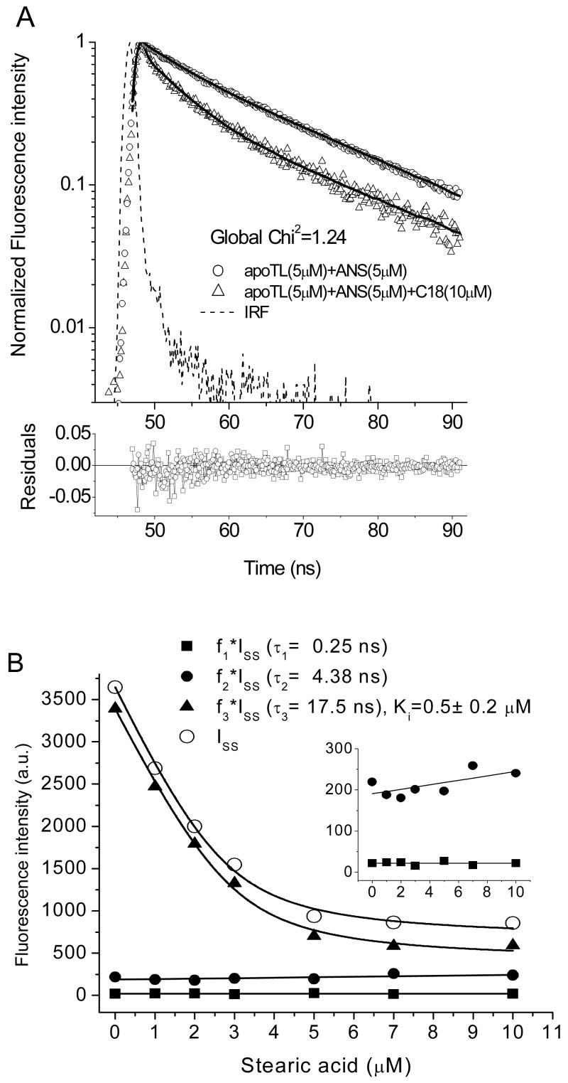
8-anilino-1-naphthalenesulfonic acid (ANS) is widely used as a probe for locating binding sites of proteins. To characterize the binding sites of tear lipocalin (TL), we studied ANS binding to apoTL by steady-state and time-resolved fluorescence. Deconvolution of ANS binding revealed that two lifetime components, 16.99ns and 2.76ns at pH 7.3, have dissociation constants of 0.58muM and 5.7muM, respectively. At pH 3.0, the lifetime components show decreased affinities with dissociation constants of 2.42muM and approximately 21muM, respectively. Selective displacement of ANS molecules from the ANS-apoTL complex by stearic acid discriminates the internal and external binding sites. Dependence of the binding affinity on ionic strength under various conditions provides strong evidence that an electrostatic interaction is involved. Time-resolved fluorescence is a promising tool to segregate multiple binding sites of proteins.


Figures

Figure 1
Figure 1
ANS fluorescence titration in the presence of apoTL (5 μM) at various buffer conditions. Solid curves are generated by fitting the experimental data to a one binding site model.
Figure 2
Figure 2
ANS fluorescence intensity decays (as an example) at various ANS concentrations with apoTL (5 μM). Solid curves are global double-decay time fit. Buffer: 10mM sodium phosphate, pH 7.3. In the figure, the number of the experimental data points has been cut in half to view clearly the differences in the ANS fluorescence intensity decays at various ANS concentrations.
Figure 3
Figure 3
Deconvolution of ANS fluorescence titration curves into lifetime components at pH 7.3 without (A) and with (B) 100 mM NaCl. The concentration of apoTL is 5 μM. Solid curves are generated by fitting of the experimental data to one binding site model.
Figure 4
Figure 4
Deconvolution of ANS fluorescence titration curves into lifetime components at pH 3.0 without (A) and with (B) 100 mM NaCl. The concentration of apoTL is 5 μM. Solid curves are generated by fitting of the experimental data to one binding site model.
Figure 5
Figure 5
Displacement of ANS form ANS (5 μM)-apoTL (5 μM) complex by stearic acid (A) ANS fluorescence intensity decays (as an example) of apoTL-ANS complex without and with stearic acid (18 μM). Solid curves are global triple-decay time fit. (B) The deconvolution of the ANS displacement curve into lifetime components. (B, inset) The free (0.24 ns) and short (4.38 ns)- lifetime components of the displacement curve. The units are same as in the main figure. Buffer: 10mM sodium phosphate, 100 mM NaCl, pH 7.3

References

    1. Flower DR. Biochem J. 1996;318(Pt 1):1–14. - PMC - PubMed
    1. Essassi D, Zini R, Tillement JP. J Pharm Sci. 1990;79:9–13. - PubMed
    1. Collini M, D'Alfonso L, Baldini G. Protein Sci. 2000;9:1968–1974. - PMC - PubMed
    1. Collini M, D'Alfonso L, Molinari H, Ragona L, Catalano M, Baldini G. Protein Sci. 2003;12:1596–1603. - PMC - PubMed
    1. D'Alfonso L, Collini M, Baldini G. Biochim Biophys Acta. 1999;1432:194–202. - PubMed

Publication types

Substances