. 2007 Nov 30;374(3):817-36.
doi: 10.1016/j.jmb.2007.08.070. Epub 2007 Sep 20.

Subunit conformations and assembly states of a DNA-translocating motor: the terminase of bacteriophage P22

Affiliations

Subunit conformations and assembly states of a DNA-translocating motor: the terminase of bacteriophage P22

Daniel Nemecek et al. J Mol Biol. .

Abstract

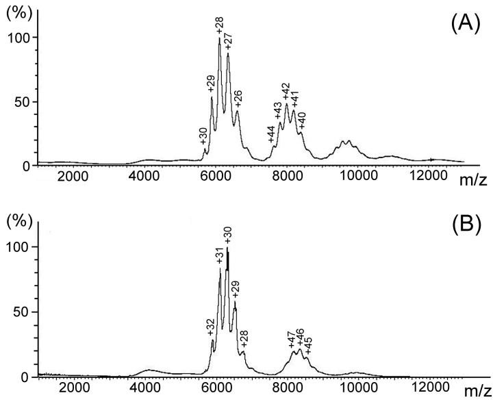
Bacteriophage P22, a podovirus infecting strains of Salmonella typhimurium, packages a 42-kbp genome using a headful mechanism. DNA translocation is accomplished by the phage terminase, a powerful molecular motor consisting of large and small subunits. Although many of the structural proteins of the P22 virion have been well characterized, little is known about the terminase subunits and their molecular mechanism of DNA translocation. We report here structural and assembly properties of ectopically expressed and highly purified terminase large and small subunits. The large subunit (gp2), which contains the nuclease and ATPase activities of terminase, exists as a stable monomer with an alpha/beta fold. The small subunit (gp3), which recognizes DNA for packaging and may regulate gp2 activity, exhibits a highly alpha-helical secondary structure and self-associates to form a stable oligomeric ring in solution. For wild-type gp3, the ring contains nine subunits, as demonstrated by hydrodynamic measurements, electron microscopy, and native mass spectrometry. We have also characterized a gp3 mutant (Ala 112-->Thr) that forms a 10-subunit ring, despite a subunit fold indistinguishable from wild type. Both the nonameric and decameric gp3 rings exhibit nonspecific DNA-binding activity, and gp2 is able to bind strongly to the DNA/gp3 complex but not to DNA alone. We propose a scheme for the roles of P22 terminase large and small subunits in the recruitment and packaging of viral DNA and discuss the model in relation to proposals for terminase-driven DNA translocation in other phages.


Figures

Figure 1
Figure 1
(A) Elution of the P22 terminase large subunit (gp2) by size exclusion chromatography (SEC). Labeled arrows near the gp2 peak maximum correspond to lanes 3-7 of the accompanying SDS-PAGE gel (inset). Also shown in the inset are molecular weight standards (lane 1) and the load fraction (lane 2). (B) Elution of the P22 terminase small subunit (gp3) by SEC. Labeled arrows near the oligomer peak correspond to lanes 2-6 and near the monomer peak to lanes 7 and 8 of the accompanying SDS-PAGE gel (inset). Also shown in the inset are molecular weight standards (lane 1).
Figure 2
Figure 2
Sedimentation velocity profile of gp2 in 0.1 M NaCl and 4 °C. Absorbances as a function of radial position were acquired at intervals of 90 s. (A) Data at intervals of 15 min (open circles) and corresponding residuals are shown for sedimentation at 42,000 rpm. The monophasic sedimentation boundaries suggest a single gp2 species. (B) Fitted distributions of the sedimentation coefficient (left) and molecular mass (right) using eqs. (1)-(4) indicate a gp2 monomer as the major species (99.6% of protein mass). The minor species (0.4%) is presumed to be a dimer.
Figure 3
Figure 3
Sedimentation equilibrium profiles of gp2 acquired at 280 nm. Samples were spun to equilibrium at speeds of 20,000 (red triangles), 25,000 (blue squares) and 30,000 (green circles) rpm in a Beckman An-Ti 60 rotor. The equilibrium profiles were fitted simultaneously by using a single-species, one-state model in Sedphat software using eq. (5).
Figure 4
Figure 4
Sedimentation velocity profiles of gp3 in 0.1 M NaCl at 4 °C. Absorbances as a function of radial position were acquired at intervals of 120 s. (A) Data at intervals of 20 min (open circles) and corresponding residuals are shown for sedimentation at 60,000 rpm. The biphasic sedimentation boundaries suggest two distinct gp3 assembly states. In this experiment the 100% monomeric gp3 sample was stored at 4°C for 24 h prior to the start of the sedimentation run. (B) Fitted distributions of the sedimentation coefficient (left) and molecular mass (right) using eqs. (1)-(4) indicate a gp3 monomer and a single multimer as the prevalent species. (C) Data as in (A) for sedimentation of 100% oligomeric gp3 at 35,000 rpm. (D) Data as in (B) for oligomeric gp3.
Figure 5
Figure 5
Sedimentation equilibrium profiles of gp3 acquired at 280, 250 and 235 nm, as labeled. Samples were spun to equilibrium at speeds of 9,000 (red triangles), 11,000 (blue squares) and 13,000 (green circles) rpm in a Beckman An-Ti 60 rotor. The equilibrium profiles were fitted simultaneously by using a single-species, two-state model in Sedphat software using eq. (5).
Figure 6
Figure 6
ESI-TOF spectra of wildtype (A) and A112T mutant (B) gp3 oligomers. Deconvolutions of the major peak clusters in the respective spectra indicate oligomer masses of 170 ± 1 and 189 ± 1 kDa, respectively, for wildtype and mutant oligomers, which correspond to nonameric and decameric assemblies. Deconvolution of the secondary cluster in each spectrum indicates a dimer of the respective oligomer. Peak labels indicate protonation states.
Figure 7
Figure 7
Electron micrographs of negatively stained wildtype (A) and A112T mutant (B) gp3 oligomers at minimal dose and 75,000X magnification. Ring morphologies of the wildtype (C) and mutant (D) oligomers revealed in two dimensions by reference-free averaging of 450 single particles in each case. The dimensions of the wildtype ring (∼10 nm outer diameter, ∼2 nm hole diameter) are about 10% smaller than those of the mutant ring. The apparent difference in intensity of the inner annuli of wildtype and mutant rings is attributed to staining artifacts.
Figure 8
Figure 8
(A) Raman spectrum (532 nm excitation) of the P22 terminase large subunit at 0.7 mg/mL in 10 mM Tris, 0.4 M NaCl. Amide I (1664 cm−1), amide III (1241 cm−1) and related markers (900-950 cm−1) indicate an α/β-fold. See Table 2. (B) Raman spectra (532 nm) of the P22 terminase small subunit [wildtype (top) and A112T (middle), each at 20 mg/mL in 10 mM Tris, 0.1 M NaCl]. Amide marker bands (1657 and 1253 cm−1) indicate α-helix as the principal secondary structure component of gp3. See Table 2. The Raman difference spectrum computed between the wildtype and mutant proteins (bottom) shows bands diagnostic of the Ala → Thr substitution and confirms the invariance of the gp3 subunit fold to the A112T mutation. The inset at upper left shows the CD profile of the gp3 monomer (open circles) and oligomer (open squares).
Figure 9
Figure 9
Native gel electromobility shift assay of gp2 and gp3 binding to 50-bp DNA. DNA and proteins are stained in green and red, respectively. (A) Robust DNA binding occurs only for monomeric (lane 3) and oligomeric (lane 5) gp3. When gp2 is added concomitantly with gp3 (lanes 4 and 6) or prior to mixing with DNA (lane 8), virtually no DNA binding is observed. Oligomeric gp3 appears to have a higher affinity than monomeric gp3 for DNA (cf. lanes 3 and 5). (B) Binding of gp2 with gp3 is demonstrated by the high stain intensity at the entry to the gel (lane 10). Minimal DNA binding is seen for free gp2 (lanes 2 and 5), while oligomeric gp3 manifests significant binding to DNA. Addition of gp2 to the DNA/gp3 complex results in the formation of a higher order (ternary) complex (lanes 4 and 7). No differences in terminase subunit binding are observed between DNAs either containing or lacking the pac site.
Figure 10
Figure 10
Proposed pathway for in vivo recognition, binding and packaging of the P22 viral genome under the action of the terminase molecular motor. Roles of the terminase large (gp2) and small (gp3) subunits in the packaging mechanism are based on the present findings and the body of previous work summarized in the review of Casjens & Weigele. Clockwise from upper left: gp3 monomer (a), or its pre-assembled ring (b), binds to concatemeric DNA to generate a complex consisting of a gp3 ring at or near the pac site. Two possible pac-site recognition schemes are depicted, viz. central annulus binding (1a), and peripheral ring binding (1b). One or several gp2 molecules are recruited to the gp3/DNA complex, resulting in cleavage of the concatemer and activation of the terminase complex, gp2/gp3/DNA (2). The activated complex docks at the procapsid portal vertex and initiates the ATPase-driven DNA packaging reaction (3). Headful packaging (∼104% of the genome length) and capsid expansion ensue (4), followed by cutting of DNA strands and release of the gp2/gp3/DNA complex from the filled capsid (5). The capsid portal is rendered competent for portal closure and tailspike attachment (not shown), while the gp2/gp3/DNA complex is reactivated (6) for another cycle of procapsid docking and headful DNA packaging (7).

Similar articles

Cited by

References

    1. Catalano CE. Viral Genome Packaging Machines: Genetics, Structure, and Mechanism. Landes Bioscience / Eurekah.com and Kluwer Academic / Plenum Publishers; Georgetown, TX and New York: 2005.
    1. Brown JC, McVoy MA, Homa FL. Packaging DNA into Herpesvirus Capsids. In: Holzenburg A, Bogner E, editors. Structure-Function Relationships of Human Pathogenic Viruses. Springer US; Cambridge, MA: 2002. pp. 111–153.
    1. Wieczorek DJ, Feiss M. Defining cosQ, the site required for termination of bacteriophage lambda DNA packaging. Genetics. 2001;158:495–506. - PMC - PubMed
    1. Casjens S, Hayden M. Analysis in vivo of the bacteriophage P22 headful nuclease. J. Mol. Biol. 1988;199:467–474. - PubMed
    1. Streisinger G, Emrich J, Stahl MM. Chromosome structure in phage T4, III. Terminal redundancy and length determination. Proc. Natl. Acad. Sci. U. S. A. 1967;57:292–295. - PMC - PubMed

Publication types

MeSH terms

LinkOut - more resources