. 2008 Jan 20;370(2):430-42.
doi: 10.1016/j.virol.2007.09.012. Epub 2007 Oct 22.

The Epstein-Barr virus BMRF-2 protein facilitates virus attachment to oral epithelial cells

Affiliations

The Epstein-Barr virus BMRF-2 protein facilitates virus attachment to oral epithelial cells

Jianqiao Xiao et al. Virology. .

Abstract

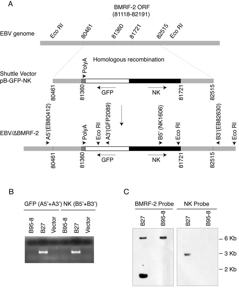
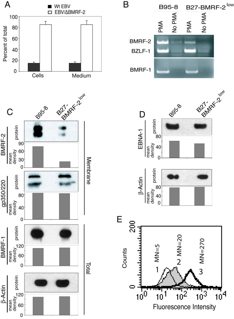
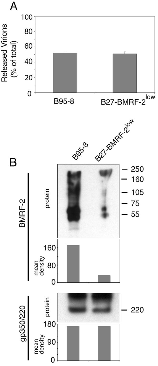
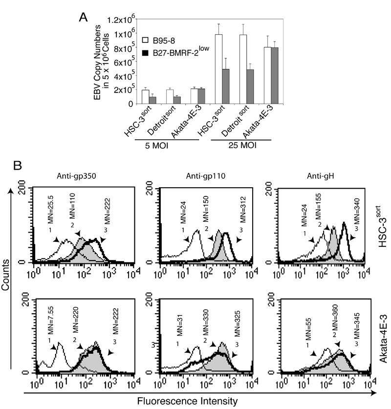
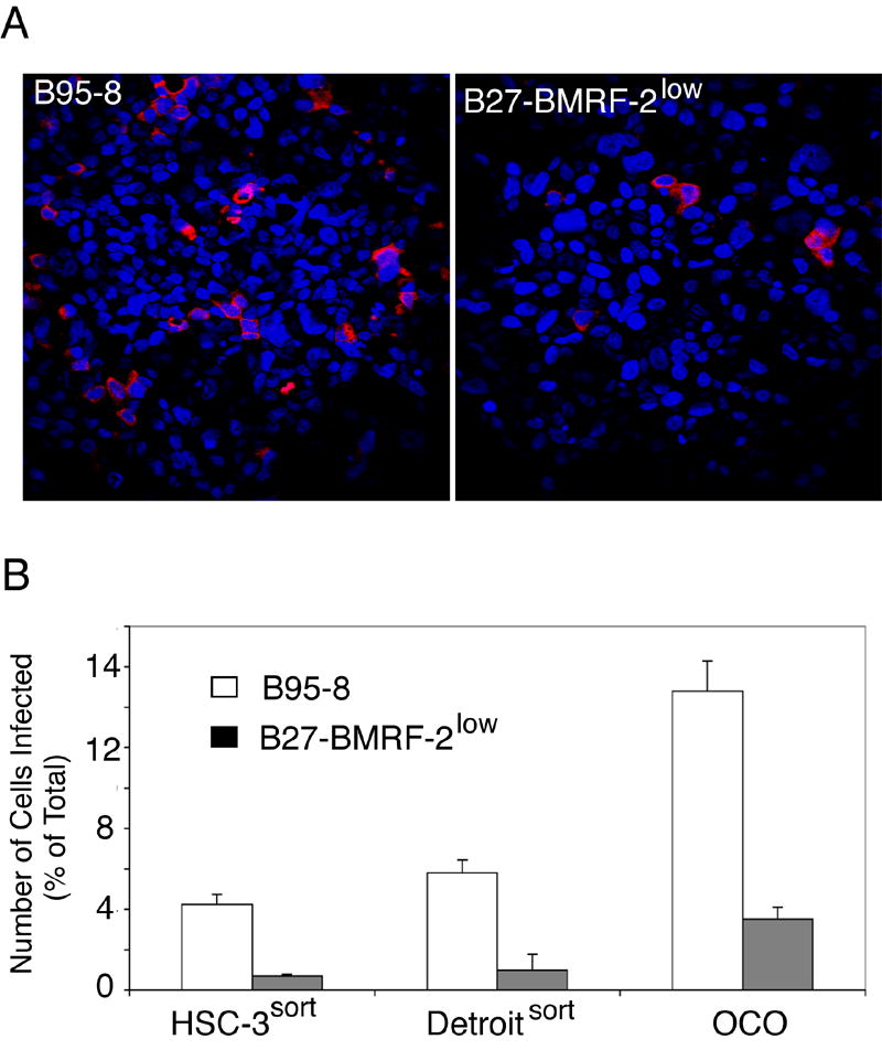
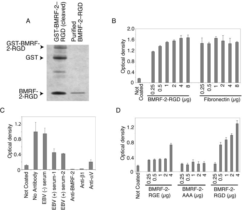
We previously reported that BMRF-2, an Epstein-Barr virus (EBV) glycoprotein, binds to beta1 family integrins and is important for EBV infection of polarized oral epithelial cells. To further study the functions of BMRF-2, we constructed a recombinant EBV that lacks BMRF-2 expression by homologous recombination in B95-8 cells. We found that lack of BMRF-2 resulted in about 50% reduction of EBV attachment to oral epithelial cells, but not to B lymphocytes, suggesting that BMRF-2 is critical for EBV infection in oral epithelial cells, but not in B lymphocytes. In polarized oral epithelial cells, infection rate of the recombinant EBV virus was about 4- to 8-fold lower than the wild-type B95-8 virus. Cell adhesion assays using the BMRF-2 RGD peptide and its RGE and AAA mutants showed that the RGD motif is critical for BMRF-2 binding to integrins. These data are consistent with our previous observation that interactions between EBV BMRF-2 and integrins are critical for infection of oral epithelial cells with EBV.


Figures

Fig. 1
Fig. 1
Construction of the recombinant EBV virus, EBV/ΔBMRF-2. A. Schematic view of the EBV genome and the shuttle vector for homologous recombination. Numbers represent the locations of the nucleotides in the EBV B95-8 genome (accession number NC_001345). B. Results of PCR using primer sets A (A5’+A3’, for GFP) and B (B5’+B3’, for NK) on genomic DNA from B95-8 and B27-BMRF-2low cells. C. Results of Southern blot analysis using a BMRF-2 probe and an NK probe and genomic DNA from B95-8 and B27-BMRF-2low cells digested with restriction enzyme Eco RI which gives a 6.4 kb (WT) band and a 1.6 kb (recombinant) band for the BMRF-2 probe, and a 3 kb (recombinant) band for the NK probe. A5’, A3’, B5’, and B3’ represents primers EB80412-31, GFP2089-2112, Nk1606-27, and EB82630-50, respectively.
Fig. 2
Fig. 2
Viral gene expression in B95-8 and B27-BMRF-2low cells. A. The ratio of recombinant EBV to WT EBV in B27-BMRF-2low cells. Total EBV copy numbers were quantitated using the BZLF-1 probe and recombinant virions were quantitated by the NK probe, numbers represent the percentages of total viral genome copy numbers. B. RT-PCR detection of BMRF-1, BMRF-2, and BZLF-1 expression in B95-8 and B27-BMRF-2low cells. 5 × 10⁶ each of B95-8 and B27-BMRF-2low cells were treated with or without PMA (30 ng/ml) for 5 days before total RNA was extracted. Five μl of cDNA (out of 20 μl reaction) were used as PCR templates. The top panel shows expression of both BMRF-2 and BZLF-1 in the same PCR reaction. The bottom panel shows expression of the BMRF-1 gene. C. Western blot analysis showing BMRF-2, gp350/220, and BMRF-1 expression in B95-8 and B27-BMRF-2low cells. For BMRF-2 and gp350 detection, 10 μg of the membrane fraction proteins were loaded onto each well of a 7 M urea SDS-PAGE gel. BMRF-1 was detected using total cell lysates separated on a NuPage 10% Bis-Tris gel. D. Western blot detection of EBNA-1 and β-actin expression in noninduced B95-8 and B27-BMRF-2low cells. 10 μg of total cell lysates were loaded onto each well in a NuPage 10% Bis-Tris gel. The mean density of pixels of the protein bands shown in panels C and D were measured by the NIH Image software, and the results of each gel are shown as a bar graph under each blot and background values were subtracted from each protein band. E. Cell surface detection of BMRF-2 by flow cytometry in B95-8 and B27-BMRF-2low cells using rat anti–BMRF-2 serum. Histograms are labeled as the following: 1, normal rat serum staining of these two cell lines (since the histograms were almost identical, only one is shown); 2, B27-BMRF-2low cells reacted with rat anti-BMRF-2 serum; 3, B95-8 cells reacted with rat anti-BMRF-2 serum. MN, mean fluorescence intensity.
Fig. 3
Fig. 3
Virion release from B95-8 and B27-BMRF-2low cells. A. Analysis of released virions by quantitative real-time PCR. B95-8 and B27-BMRF-2low cells were cultured in the presence of PMA and butyric acid for 5 days, and total genomic DNA from cells or media was extracted separately. EBV genome copy numbers were quantitated by real-time PCR and number of released virions in medium from B95-8 or B27-BMRF-2low cells were shown as % of total, which equals to the number of virions from cells and medium together. Error bars represent standard deviations (SD) of three independent experiments. B. Western blot analysis of double gradient purified virions from B95-8 or B27-BMRF-2low cells using rat anti–BMRF-2 serum. 10 μg of total viral proteins were loaded onto each well of a 7 M urea SDS-PAGE gel.
Fig. 4
Fig. 4
Analysis of attachment of B95-8 and B27-BMRF-2low virus to oral epithelial and Akata 4E-3 B lymphocytes. A. Detection of virus attachment by real-time PCR. The B95-8 and B27-BMRF-2low viruses were added independently to HSC-3sort, Detroitsort, and Akata 4E-3 cells and incubated at 4°C for 1 h. Cells were washed, and genomic DNA extracted and bound virions were examined by real-time PCR. B. Evaluation of virus binding by flow cytometry assay. HSC-3sort and Akata 4E-3 cells were incubated with B95-8 or B27-BMRF-2low viruses for 1 h at 4°C and washed, and attached virions were detected using mouse monoclonal anti-gp250/350/ (left panel), anti-gp110 (anti-gB, middle panel), or anti-gH (right panel). The amount of virions bound to the cell surface was examined by flow cytometry and the results are shown as histograms labeled as follows: 1, cells incubated with the B95-8 virus and reacted with mouse IgG1; 2 and 3, cells incubated with the B27-BMRF-2low and the B95-8 virus, respectively, and reacted with one of the above monoclonal antibodies. MN, mean fluorescence intensity.
Fig. 5
Fig. 5
Infection of polarized oral epithelial cells with B95-8 or B27-BMRF-2low virus. A. Polarized OCO cells were infected with B95-8 or B27-BMRF-2low virus from their basolateral membranes at 100 MOI. At 5 days after infection, cells were fixed and stained with mouse anti-gp350/220 antibody (red). Cell nuclei were stained in blue. B. Polarized HSC-3sort, Detroitsort and OCO cells were infected with B95-8 or B27-BMRF-2low virus from their basolateral membranes at 100 MOI/cell. Cells were fixed at 5 days postinfection and immunostained with gp350/220 antibodies; positive cells were counted. Numbers shown are the average percentage of gp350/220–positive cells from individual filer membranes. Error bars show SD (n=3).
Fig. 6
Fig. 6
Adhesion of HSC-3sort cells to BMRF-2 RGD–coated plates. A. Purification of the BMRF-2 RGD peptide: BMRF-2 RGD fragment was cleaved by thrombin from the GST-BMRF-2 RGD fusion protein and purified by Centricon-10. Ten μg of GST-BMRF-2 RGD fusion protein (left lane) and 1 μg of purified BMRF-2 RGD peptide (right lane) were separated on an SDS PAGE gel and stained by simple blue (Invitrogen). B. Adhesion of HSC-3sort cells to a Maxisorp 96-well plate coated with the BMRF-2 RGD peptide or fibronectin at different concentrations. C. Adhesion of HSC-3sort cells was blocked by different antibodies: EBV-negative serum, EBV-positive serum, or rat anti-BMRF-2 were added to a Maxisorp 96-well plate coated with the BMRF-2 RGD peptide before HSC-3sort cells were applied to the plate; Anti-β1 and anti-αv antibodies were incubated with HSC-3sort cells on ice for 60 min before they were added to the BMRF-2 RGD–coated plate. D. Adhesion of HSC-3sort cells to the wells of a Maxisorp 96-well plate coated with PBS, BMRF-2 RGD peptide, BMRF-2 RGE peptide, or BMRF-2 AAA peptide. B-D, Cell adhesion was examined by measuring attached cells after crystal violet staining using an ELISA-plate reader with a 595 nm absorbance filter. Error bars represent SD of three independent experiments.

References

    1. Adam T. Exploitation of host factors for efficient infection by Shigella. Int J Med Microbiol. 2001;291(4):287–98. - PubMed
    1. Akula SM, Pramod NP, Wang FZ, Chandran B. Integrin alpha3beta1 (CD 49c/29) is a cellular receptor for Kaposi’s sarcoma-associated herpesvirus (KSHV/HHV-8) entry into the target cells. Cell. 2002;108(3):407–19. - PubMed
    1. Borza CM, Morgan AJ, Turk SM, Hutt-Fletcher LM. Use of gHgL for attachment of Epstein-Barr virus to epithelial cells compromises infection. J Virol. 2004;78(10):5007–14. - PMC - PubMed
    1. Chu JJ, Ng ML. Interaction of West Nile virus with alpha v beta 3 integrin mediates virus entry into cells. J Biol Chem. 2004;279(52):54533–41. - PubMed
    1. Ciarlet M, Crawford SE, Cheng E, Blutt SE, Rice DA, Bergelson JM, Estes MK. VLA-2 (alpha2beta1) integrin promotes rotavirus entry into cells but is not necessary for rotavirus attachment. J Virol. 2002;76(3):1109–23. - PMC - PubMed

Publication types

MeSH terms