Review
. 2007 Oct;19(5):521-8.
doi: 10.1016/j.ceb.2007.09.001. Epub 2007 Oct 22.

Gap junction channel structure in the early 21st century: facts and fantasies

Affiliations
Review

Gap junction channel structure in the early 21st century: facts and fantasies

Mark Yeager et al. Curr Opin Cell Biol. 2007 Oct.

Abstract

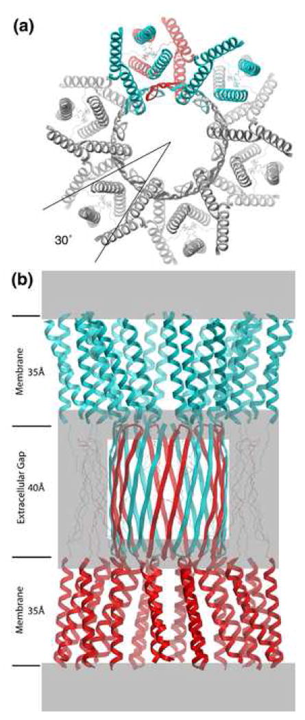
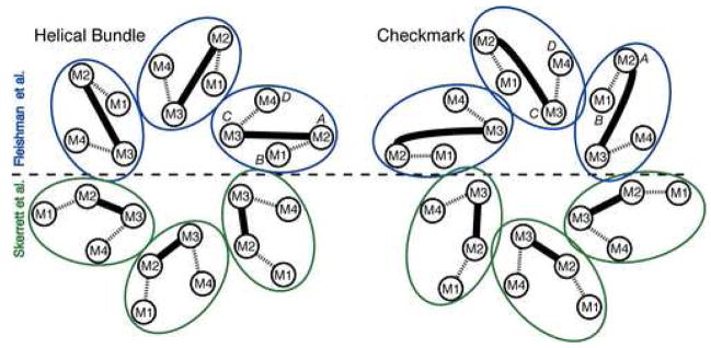
Gap junction channels connect the cytoplasms of adjacent cells through the end-to-end docking of single-membrane structures called connexons, formed by a ring of six connexin monomers. Each monomer contains four transmembrane alpha-helices, for a total of 24 alpha-helices in a connexon. The fundamental structure of the connexon pore is probably similar in unpaired connexons and junctional channels, and for channels formed by different connexin isoforms. Nevertheless, variability in results from structurally focused mutagenesis and electrophysiological studies raise uncertainty about the specific assignments of the transmembrane helices. Mapping of human mutations onto a suggested C(alpha) model predicts that mutations that disrupt helix-helix packing impair channel function. An experimentally determined structure at atomic resolution will be essential to confirm and resolve these concepts.


Figures

Figure 1
Figure 1
Molecular design of gap junction channels. (a) Top view showing the 30 ° rotational stagger between docked connexons. Two subunits of the top connexon (in blue) are above one subunit of the bottom connexon (in red). The other subunits have been colored gray for clarity. The molecular boundary is depicted as a 4-helix bundle, but there are other possibilities (Figure 2). (b) Side view. The top connexon is in blue and the bottom one in red. Grayed areas denote parts of the structure that are most uncertain, especially the folding within the density at the boundary between the transmembrane assembly and the extracellular space. Putative β sheets corresponding to E1 (on the perimeter of the extracellular gap) are drawn with thin lines to emphasize this ambiguity. The E2 loops are depicted as an interdigitating β barrel [51]. Refer to Figures 2 and 3 in reference [7] for the corresponding views of the 3D density map derived by electron cryo-crystallography. The dimensions for the transmembrane helical domain and the extracellular gap are approximate. From [22], reproduced by permission.
Figure 2
Figure 2
Possible molecular boundaries for the connexin subunit include a helical bundle (left) and a “checkmark” (right) (following the naming of Unger et al. [7]). Each shows two assignments for the 4 transmembrane α-helices within each subunit, according to Fleishman et al. [8] (top, blue) and Skerrett et al. [43] (bottom, green). Dashed lines denote the extracellular loops, E1 and E2, and the solid lines denote the M2-M3 cytoplasmic loops. The α-helical rods designated A, B, C and D in the 3D density map derived by electron cryo-crystallography (Unger et al. [7] are also indicated. Both assignments shown designate M3 as the major pore lining helix, but other studies suggest M1, as discussed in the text. From [22], reproduced by permission.
Figure 3
Figure 3
A Cα model (yellow ribbons) for the membrane spanning α-helices of a connexon, derived by combining the information from a computational analysis of connexin sequences, the results of a number of biochemical studies, and the constraints provided by a 3D cryo-EM map (blue) [8]. While individually, none of these approaches provided high-resolution information, their sum yielded an atomic model that predicts how connexin mutations (red spheres) that result in diseases such as nonsyndromic deafness and Charcot-Marie-Tooth disease may interfere with formation of functional channels by disrupting helix-helix packing. Adapted from [8].
Figure 4
Figure 4
CryoEM structure of two-dimensional crystals of reconstituted Cx26 connexons. The 3D map is contoured at 1σ (light blue) and 2.4 σ (yellow) above the mean density. The inset in the upper left shows a 20-Å-thick section perpendicular to the membrane plane through the density map of a connexon. This section corresponds to the region enclosed by the white lines shown in A. The arrowhead points to the large plug of density within the pore. The inner cytoplasmic protrusions (white arrows) extend from the cytoplasmic ends of helices B and C. (AC). 30-Å-thick slabs through the density map corresponding to the position of the lines shown in the Inset. The four α-helices are labeled A (cyan, A′), B (green, B′), C (yellow), and D (pink) as in the original Cx43 structure (Figure 2) [7]. The arrowhead and white arrows represent the plug and the inner cytoplasmic protrusions, respectively, as in Inset. From [9], reproduced by permission.

Similar articles

Cited by

References

    1. Loewenstein WR. Junctional intercellular communication: the cell-to-cell membrane channel. Physiol Rev. 1981;61:829–913. - PubMed
    1. Bruzzone R, White TW, Paul DL. Connections with connexins: the molecular basis of direct intercellular signaling. Eur J Biochem. 1996;238:1–27. - PubMed
    1. Gerido DA, White TW. Connexin disorders of the ear, skin, and lens. Biochim Biophys Acta. 2004;1662:159–170. - PubMed
    1. Unwin PN, Ennis PD. Two configurations of a channel-forming membrane protein. Nature. 1984;307:609–613. - PubMed
    1. Unger VM, Kumar NM, Gilula NB, Yeager M. Projection structure of a gap junction membrane channel at 7 A resolution. Nat Struct Biol. 1997;4:39–43. - PubMed

Publication types