. 2007 Dec 21;282(51):37244-55.
doi: 10.1074/jbc.M708137200. Epub 2007 Oct 17.

Control of phenotypic plasticity of smooth muscle cells by bone morphogenetic protein signaling through the myocardin-related transcription factors

Affiliations

Control of phenotypic plasticity of smooth muscle cells by bone morphogenetic protein signaling through the myocardin-related transcription factors

Giorgio Lagna et al. J Biol Chem. .

Abstract

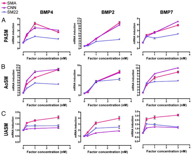
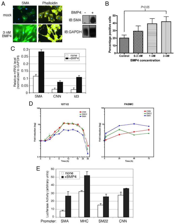
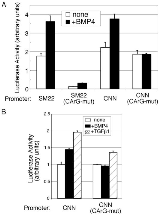
Vascular smooth muscle cells (VSMCs), unlike other muscle cells, do not terminally differentiate. In response to injury, VSMCs change phenotype, proliferate, and migrate as part of the repair process. Dysregulation of this plasticity program contributes to the pathogenesis of several vascular disorders, such as atherosclerosis, restenosis, and hypertension. The discovery of mutations in the gene encoding BMPRII, the type II subunit of the receptor for bone morphogenetic proteins (BMPs), in patients with pulmonary arterial hypertension (PAH) provided an indication that BMP signaling may affect the homeostasis of VSMCs and their phenotype modulation. Here we report that BMP signaling potently induces SMC-specific genes in pluripotent cells and prevents dedifferentiation of arterial SMCs. The BMP-induced phenotype switch requires intact RhoA/ROCK signaling but is not blocked by inhibitors of the TGFbeta and PI3K/Akt pathways. Furthermore, nuclear localization and recruitment of the myocardin-related transcription factors (MRTF-A and MRTF-B) to a smooth muscle alpha-actin promoter is observed in response to BMP treatment. Thus, BMP signaling modulates VSMC phenotype via cross-talk with the RhoA/MRTFs pathway, and may contribute to the development of the pathological characteristics observed in patients with PAH and other obliterative vascular diseases.


Figures

FIGURE 1
FIGURE 1. BMP pathway prevents dedifferentiation of human primary PASMCs
A, confluent hPAMSCs (passage 5) were cultured in growth media containing 3 nM BMP4 or vehicle. Total RNAs were collected at 0, 5, and 8 days after the treatment and were subjected to RT-PCR analysis of human SMA and GAPDH (loading control). B, hPASMCs at passage 7 (high passage) or passage 4 (low passage) were treated with 3 nM BMP4 or 400 pM TGFβ1 for 72 h in DMEM/0.1% FCS. Cells were then subjected to immunofluorescence staining with FITC-conjugated anti-SMA antibody (green) and nuclear staining with DAPI (blue). C, hPASMCs (passage 5) were treated with 20 ng/ml PDGF-BB and/or 3 nM BMP4 or 400 pM TGFβ1 for 72 h in DMEM/0.1% FCS. Cells were then subjected to immunofluorescence staining with anti-SMA antibodies and nuclear staining with DAPI. D, rat PAC-1 cells were treated with 3 nM BMP4 and/or 100 pM TGFβ1 for 48 h in DMEM/10% FCS. Cells were subjected to immunofluorescence staining with FITC-conjugated anti-SMA antibodies (green) and nuclear staining with DAPI (blue). E, hPASMCs at passage 5 were treated with 20 ng/ml PDGF-BB and/or 3 nM BMP4 or 400 pM TGFβ1 for 72 h in DMEM/0.1% FCS. Total RNAs were prepared from the cells and subjected to semi-quantitative RT-PCR analysis of SMA and GAPDH (control). Results were normalized to GAPDH expression. F, hPASMCs at passage 5 were treated with 20 ng/ml PDGF-BB and/or 3 nM BMP4 or infection with recombinant adenovirus carrying HA epitope-tagged constitutively active (CA) ALK6 (m.o.i 200). Cells were subjected to immunofluorescence stain with FITC-conjugated anti-SMA antibody (green) and anti-HA antibodies conjugated with Alexa Fluor 555 (for ALK6; yellow) and nuclear stain with DAPI (blue) (top panels). Total cell lysates prepared from hPASMCs treated with or without 3 nM BMP4 for 48 h were subjected to immunoblot with anti-SMA antibody or GAPDH (loading control) (bottom panel). G, hPASMCs at passage 5 were infected with recombinant adenovirus expressing HA-tagged dominant negative (DN) ALK2, ALK3, ALK6, or BMPRII (m.o.i. 200). Cells were subjected to immunofluorescence stain with anti-SMA (green) and anti-HA antibody (for ALK2, 3, 6, and BMPRII; yellow) and nuclear stain with DAPI (blue).
FIGURE 2
FIGURE 2. Induction of SM-markers by BMPs in different vascular SMCs
Three types of vascular SMCs, hPASMC (A), hAoSM (B), and hUASM (C), were treated with BMP2, BMP4, or BMP7 at different concentrations (0.3–3 nM) in DMEM/0.2% FCS for 24 h. Total RNA was isolated and subjected to RT-PCR analysis using primers for SMA, calponin (CNN), and SM22α (SM22). Relative mRNA expression of each gene was normalized to GAPDH mRNA.
FIGURE 3
FIGURE 3. BMP4 signaling induces SM gene expression in a Smad-dependent manner
hPASMCs were transfected with a mixture of siRNA against Smad1 and Smad5 (siSmads) or control siRNA (siScr) for 24 h prior to treatment with 3 nM BMP4 for 48 h. Total RNA was isolated and subjected to RT-PCR analysis using primers for SMA, calponin, and the non-SM-specific, BMP-target gene Id3. Relative mRNA expression of each gene was normalized to GAPDH mRNA. The mRNA expression of Smad1 or Smad5 indicates reduction by siRNA.
FIGURE 4
FIGURE 4. BMP4 induces expression of SMA in non-SMCs
A, 10T½ cells were treated with 3 nM BMP4 for 72 h in DMEM/10% FCS. Cells were subjected to immunofluorescence staining with FITC-conjugated anti-SMA antibodies (green), Alexa Fluor 568-conjugated phalloidin (yellow), and nuclear staining with DAPI (blue) (left panels). Total cell lysates prepared from 10T½ cells treated with 3 nM BMP4 or vehicle for 48 h were subjected to immunoblot with anti-SMA antibody (right panel) or GAPDH (loading control). B, 10T½ cells were treated with 3 nM BMP4 at 0.3, 1, or 3 nM and subjected to immunofluorescence staining with FITC-conjugated anti-SMA antibodies. SMA-positive cells were counted and are shown as percentage of total cells (n = 100). The increase in number of SMA-positive cells observed upon treatment with 3 nM BMP4 is statistically significant compared with control cells (Student’s t test, p < 0.05). C, 10T½ cells were treated with 3 nM BMP4 in DMEM/0.2% FCS for 24 h. Total RNA was isolated and subjected to RT-PCR analysis using primers for SMA, calponin, and non-SM, BMP4-target gene Id3. Relative mRNA expression of each gene was normalized to GAPDH mRNA. D, 10T½ cells were treated with 3 nM BMP4 for various lengths of time (2– 48 h). Total RNA was isolated and subjected to RT-PCR analysis using primers for SMA, calponin (CNN), and SM22α (SM22). Relative mRNA expression of each gene was normalized to GAPDH mRNA. E, different SM gene promoter (SMA, CNN, SM-MHC, and SM22α)-luciferase reporter constructs were transiently transfected into 10T½ cells as indicated. Cells were treated with or without 3 nM BMP4 for 20 h, followed by luciferase assay.
FIGURE 5
FIGURE 5. Involvement of the RhoA/ROCK pathway in the regulation of SMA by BMP4
A, hPASMCs were treated with specific inhibitor of the type I TGFβ receptor kinase (SB-431542) prior to 3 nM BMP4 or 100 pM TGFβ1 stimulation, followed by immunostaining with FITC-conjugated anti-SMA antibody (green) and nuclear stain with DAPI (blue). B, 10T½ cells were transfected with the SMA-luciferase reporter. Cells were infected with adenovirus expressing constitutively active RhoA [RhoA (CA)], dominant negative RhoA [RhoA (DN)] or vehicle for 24 h, then treated with 3 nM BMP4 for 48 h with or without 10 μM ROCK inhibitor (Y-27632) and assayed for luciferase activity. C, hPASMCs were treated with increasing concentrations (100 nM–10 μM) of PI-3K inhibitor (Wortmannin) or ROCK inhibitor (Y-27632), with or without 3 nM BMP4 for 48 h, followed by immunostaining with FITC-conjugated anti-SMA antibody (green) and nuclear stain with DAPI (blue).
FIGURE 6
FIGURE 6. BMP-mediated transcriptional activation of SM gene is CArG box-dependent
A, 10T½ cells were transfected with wild-type or mutant SM22α or calponin reporter constructs, which are mutated in the CArG box sequence as indicated, treated with BMP4, and assayed for luciferase activity. B, PAC-1 cells were transfected with wild-type or mutant calponin reporter constructs, which are mutated in the CArG box sequence as indicated, treated with 3 nM BMP4 or 100 pM TGFβ1 and assayed for luciferase activity.
FIGURE 7
FIGURE 7. BMP4 signal mediatesarecruitmentofcofactorMRTFstoSMApromoter
A,10T½ cellsweretransfected with SMA-reporter constructs and increasing amounts of Myc-MRTF-A expression plasmid (1, 2, 3, 4, or 5 ng) and assayed for luciferase activity. B, total RNAs were extracted from 10T½ cells transfected with siRNA against MRTF-A or MRTF-B or non-targeting (control) siRNA and were subjected to RT-PCR analysis to examine expression of MRTF-A or MRTF-B mRNA. RNA levels were quantified by real-time PCR analysis with normalization to GAPDH expression. C, 10T½ cells transfected with MRTF-A or MRTF-B siRNAs, or non-targeting (control) siRNA, together with the SMA reporter construct. The luciferase activity was measured after treating cells with or without 3 nM BMP4 for 48 h. There is no statistically significant change in the SMA reporter activity in cells transfected with MRTF-A siRNA, unlike cells transfected with control siRNA or MRTF-B siRNA (Student’s t test, n.s. represents p > 0.05). D, 10T½ cells transfected with siRNAs as in panel C, and treated with or without 3 nM BMP4 or 400 pM TGFα1, followed by immunostaining with FITC-conjugated anti-SMA antibody (green) and nuclear stain with DAPI (blue). As negative control, non-targeting siRNA was used. E, 10T½ cells were transiently transfected with Myc-MRTF-A expression plasmid. After being treated with 3 nM BMP4, 20% serum, or BMP4 and Latrunculin B (LB), an inhibitor of actin polymerization, cells were subjected to immunostaining with FITC-conjugated anti-Myc antibody (green) and nuclear stain with DAPI (blue). The cells in which MRTF-A localization was predominantly nuclear were counted and compared as percentage to the total number of cells. F, 10T½ cells were transfected with Myc-tagged myocardin-856 (smooth muscle isoform), MRTF-A, or MRTF-B and treated with 3 nM BMP4 for 24 h. Cells were then subjected to ChIP assay. A recruitment of these cofactors to the SMA promoter was examined by immunoprecipitation with an anti-Myc antibody, followed by quantitative real time-PCR analysis using primers specific for the SMA promoter. As control, untransfected cells were subjected to the ChIP assay using anti-Myc or anti-SRF antibody, followed by PCR using the same primers. An average of triplicate experiments is presented. The data show induction of SMA chromatin binding by BMP4 compared with cells treated with vehicle. In the inset, data are normalized to the background signal observed with the anti-Myc antibody immunoprecipitation in untransfected cells. The amount plotted in the input bar is 1:60 of the total used for each immunoprecipitation.

Similar articles

Cited by

References

    1. Owens G. Physiol Rev. 1995;75:487–517. - PubMed
    1. Rubin LJ, Rich S. Primary Pulmonary Hypertension. Marcel Dekker, Inc; New York, NY: 1997.
    1. Runo JR, Loyd JE. Lancet. 2003;361:1533–1544. - PubMed
    1. Machado RD, Pauciulo MW, Thomson JR, Lane KB, Morgan NV, Wheeler L, Phillips JA, 3rd, Newman J, Williams D, Galie N, Manes A, McNeil K, Yacoub M, Mikhail G, Rogers P, Corris P, Humbert M, Donnai D, Martensson G, Tranebjaerg L, Loyd JE, Trembath RC, Nichols WC. Am J Hum Genet. 2001;68:92–102. - PMC - PubMed
    1. Thomson JR, Machado RD, Pauciulo MW, Morgan NV, Humbert M, Elliott GC, Ward K, Yacoub M, Mikhail G, Rogers P, Newman J, Wheeler L, Higenbottam T, Gibbs JS, Egan J, Crozier A, Peacock A, Allcock R, Corris P, Loyd JE, Trembath RC, Nichols WC. J Med Genet. 2000;37:741–745. - PMC - PubMed

Publication types

MeSH terms