

Gadolinium-enhanced MR cisternography to evaluate dural leaks in intracranial hypotension syndrome
- PMID: 17947371
- PMCID: PMC8119121
- DOI: 10.3174/ajnr.A0746
Gadolinium-enhanced MR cisternography to evaluate dural leaks in intracranial hypotension syndrome
Abstract
Background and purpose: We evaluated the use of MR cisternography after intrathecal administration of gadopentetate dimeglumine to detect the presence and localization of CSF leaks in 19 patients diagnosed with spontaneous intracranial hypotension syndrome according to the criteria of International Headache Society.
Materials and methods: Lumbar puncture with an injection of 0.5 mL of gadopentetate dimeglumine into the subarachnoid space in the lumbar area was performed. MR images of the cervical, thoracic, and lumbar regions in axial, coronal, and sagittal planes with fat-saturated T1-weighted images were acquired.
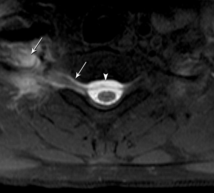
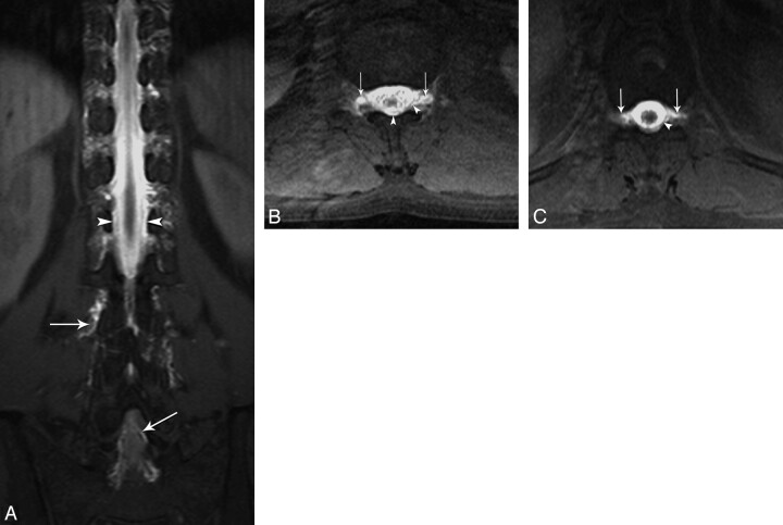
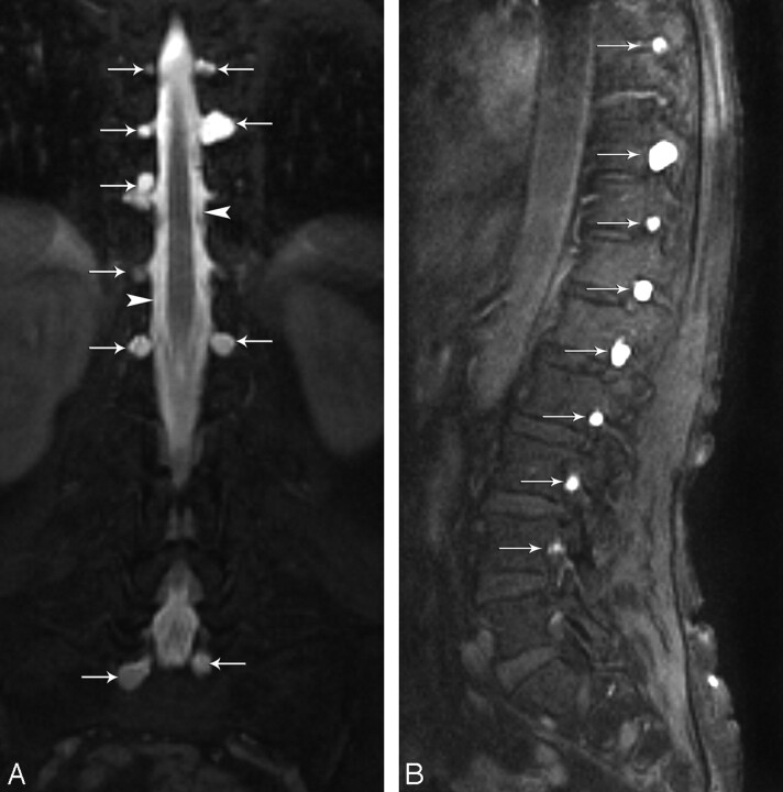
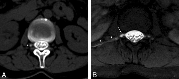
Results: We observed objective CSF leakage in 17 (89%) of 19 patients. In 14 of these 17 patients, the site of dural tear was demonstrated accurately. In 3 of these 17 patients, the contrast leakage was diffuse, and site of the leak could not be located accurately. No leakage was observed in 2 patients. No complications were detected in any of the patients during the first 24 hours after the procedure or during the 6- to 12-month follow-up.
Conclusion: The current results demonstrate the relative safety, accuracy, and feasibility of intrathecal gadolinium-enhanced MR cisternography to evaluate dural leaks.
Figures
Comment in
-
Intrathecal gadolinium: its time has come?AJNR Am J Neuroradiol. 2008 Jan;29(1):3-4. doi: 10.3174/ajnr.A0884. AJNR Am J Neuroradiol. 2008. PMID: 18192343 Free PMC article. No abstract available.
References
-
- Schaltenbrand G. Neuere anschauungen zur pathophysiologie der liquorzirkulation. Zentralbl Neurochir 1938;3:290–300
-
- Schaltenbrand G. Normal and pathological physiology of the cerebrospinal fluid circulation. Lancet 1953;1:805–08 - PubMed
-
- Headache Classification Subcommittee of the International Headache Society. The international classification of headache disorders: 2nd ed. Cephalalgia 2004;24(suppl 1):9–160 - PubMed
-
- Mokri B, Krueger BR, Miller GM, et al. Meningeal gadolinium enhancement in low-pressure headaches. Ann Neurol 1991;30:294–95
-
- Mokri B. Spontaneous cerebrospinal fluid leaks: from intracranial hypotension to cerebrospinal fluid hypovolemia–evolution of a concept [review]. Mayo Clin Proc 1999;74:1113–23 - PubMed
MeSH terms
Substances
LinkOut - more resources
Full Text Sources
Medical