Comparative Study
. 2007 Nov 14;26(22):4732-43.
doi: 10.1038/sj.emboj.7601885. Epub 2007 Oct 18.

Spinocerebellar ataxia with axonal neuropathy: consequence of a Tdp1 recessive neomorphic mutation?

Affiliations
Comparative Study

Spinocerebellar ataxia with axonal neuropathy: consequence of a Tdp1 recessive neomorphic mutation?

Ryuki Hirano et al. EMBO J. .

Abstract

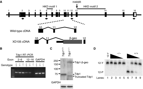
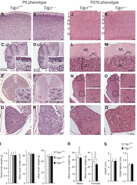
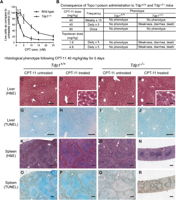
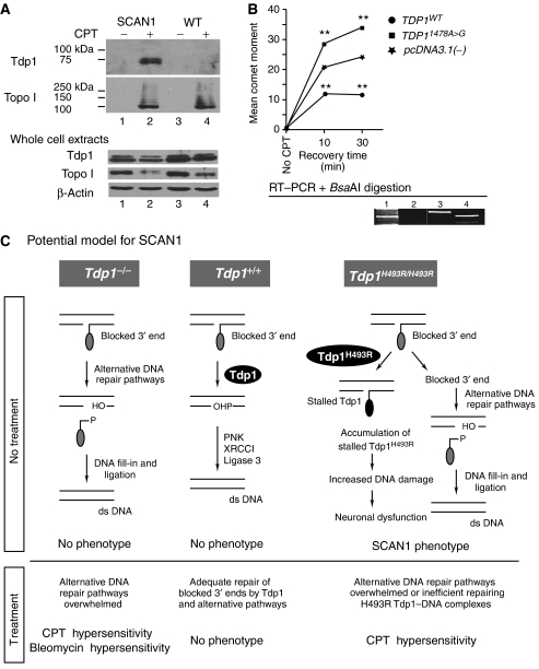
Tyrosyl-DNA phosphodiesterase 1 (Tdp1) cleaves the phosphodiester bond between a covalently stalled topoisomerase I (Topo I) and the 3' end of DNA. Stalling of Topo I at DNA strand breaks is induced by endogenous DNA damage and the Topo I-specific anticancer drug camptothecin (CPT). The H493R mutation of Tdp1 causes the neurodegenerative disorder spinocerebellar ataxia with axonal neuropathy (SCAN1). Contrary to the hypothesis that SCAN1 arises from catalytically inactive Tdp1, Tdp1-/- mice are indistinguishable from wild-type mice, physically, histologically, behaviorally, and electrophysiologically. However, compared to wild-type mice, Tdp1-/- mice are hypersensitive to CPT and bleomycin but not to etoposide. Consistent with earlier in vitro studies, we show that the H493R Tdp1 mutant protein retains residual activity and becomes covalently trapped on the DNA after CPT treatment of SCAN1 cells. This result provides a direct demonstration that Tdp1 repairs Topo I covalent lesions in vivo and suggests that SCAN1 arises from the recessive neomorphic mutation H493R. This is a novel mechanism for disease since neomorphic mutations are generally dominant.


Figures

Figure 1
Figure 1
Generation of the Tdp1−/− mouse by insertional mutagenesis. (A) Diagram of the mouse Tdp1 genomic structure (top). The H493R mutation found in SCAN1 patients would be encoded in exon 13 of mouse Tdp1. For generation of the Tdp1-deficient mice (Tdp1−/−), we used embryonic stem cells containing a pGT1Lxf insertion in Tdp1 intron 11. This insertion truncates the Tdp1 mRNA following exon 11. The location of the primers used for genotyping and RT–PCR detection are indicated. (B) RT–PCR confirmation of the absence of full-length Tdp1 mRNA expression in the brain of Tdp1−/− mice. The upstream RT–PCR primers reside in exons 2 and 6, the downstream primers in exons 12 and 16. (C) Western confirmation of the absence of expression of full-length Tdp1 protein in Tdp1−/− neurospheres. The western analysis also detected the Tdp1-β-geo fusion protein and a smaller Tdp1 product that is the product of alternative splicing deleting exons 12 and 13. (D) Absence of Tdp1 enzymatic activity in lysates from Tdp1−/− mouse neurospheres as shown by the failure to hydrolyze the tyrosyl-DNA substrate 12-Y. Each lane represents a serial 10-fold dilution of the Tdp1−/− (lanes 2–5) and Tdp1+/+ (lanes 6–9) cell extracts.
Figure 2
Figure 2
Comparative phenotypic and histopathological evaluation of the Tdp1+/+ and Tdp1−/− mice at P0 (A–I) and P270 (J–S). P0 mice: Hematoxylin and eosin (H&E) staining of cortex (A, B), cerebellar vermis (C, D), spinal cord (E, F), and dorsal root ganglion (G, H). Note the comparability of the external granular cell (EGL), molecular (ML), and internal granular cell layers (IGL) as well as the Purkinje cell number (PC). (I) Comparison of the body weights and lengths and brain weights of Tdp1+/+, Tdp1+/−, and Tdp1−/− littermates. P270 mice: H&E staining of cortex (J, K), cerebellar vermis (L, M), spinal cord (N, O), and dorsal root ganglia (P, Q). The cortical layers are labeled in panels J and K and the cerebellar molecular (ML) and granular cell (GL) layers in panels L and M. The insets in panels N and O are higher magnifications of the dorsal (upper) and ventral (lower) horns. (R) Comparison of male and female body weights. (S) Comparison of the peripheral nerve electrophysiology of Tdp1+/+ and Tdp1−/− littermates. Scale bar=50 μm.
Figure 3
Figure 3
Immunohistochemical analysis of Tdp1 protein expression in the human brain. (A) Tdp1 expression in the cerebrum of a human 10 gestational week (GW) brain. Cells adjoining the ventricle (V), and in the subventricular zone (SVZ), intermediate zone (IMZ), and cortical plate (CP) express Tdp1. (B, C) Tdp1 expression in layer two of the cortex from a 40 GW and a 16-year brain. (DF) Tdp1 expression in the cerebella from a 22 GW, a 9-month and a 4-year brain. Note the staining in the external granular cell layer (EGL), Purkinje cells (PC), and internal granular cell layer (IGL). (G) Tdp1 expression in a 19-year dentate gyrus. (H, I) Tdp1 expression in spinal cord anterior horn cells (AHC) and a dorsal root ganglion of a 10-year old, respectively. (JM) Specificity of the immune serum is shown by absence of staining of the spinal cord (J) and dorsal root ganglion (K) from a 10-year old with preimmune serum and also by absence of antigen recognition in mouse Tdp1−/− mouse embryonic fibroblasts compared to Tdp1−/− fibroblasts transfected with a human Tdp1 expression construct (Tdp1−/−+pCMV5·Tdp1) (L), and competitive inhibition of the antiserum by recombinant human Tdp1 (M). Scale bar=50 μm.
Figure 4
Figure 4
In situ hybridization and immunohistochemistry showing the spatial and temporal expression of mouse Tdp1 mRNA and protein, respectively. Localization of the Tdp1 mRNA in P1 (AD), P10 (EH), and adult (IL) mice. (A, E, I) Expression of Tdp1 mRNA in a midline sagittal section from a P1, P10, and adult mouse or brain, respectively. Note that Tdp1 is expressed throughout the cortex (Cx), cerebellum (Cb), spinal cord (SC), and dorsal root ganglia (DRG) as well as most other organs including the skin, thymus (Th), heart, lungs, liver, and intestines. (B, F, J) Expression of Tdp1 mRNA in the cortex (Cx) and hippocampus (Hip) of a P1, P10, and adult brain, respectively. (C, G, K) Expression of Tdp1 mRNA in a coronal section of a P1, P10, and adult cervical spinal cord, respectively. The highest expression is in the dorsal horns (DH) and the ventral horns (VH). (D, H, L) Expression of Tdp1 mRNA in a cross-section of a P1, P10, and adult dorsal root ganglion, respectively. (MO) Serial sections from the P1 mouse hybridized with a sense probe for Tdp1 showing the specificity of the hybridization for the antisense probe used in panels A–L. Scale bar=1 cm (A, E, I, M), 0.5 mm (B, C, F, G, J, K, N, O), 0.2 mm (D, H, L). Localization of the Tdp1 protein in the P0 and the P270 CNS (P-AA). (P) P0 cortex and hippocampus, (Q) P0 layer II cortical neurons, (R) P270 cortex, (S) P270 layer II cortical neurons, (T) P0 cerebellum, (U) P270 cerebellum, (V) P270 dentate nucleus, (W) P270 spinal cord. Nonspecific staining of the P0 cortex (X), P270 cortex (Y), P0 cerebellum (Z), and P270 cerebellum (AA). Scale bar=0.5 mm (P, R, T, U, Y, Z, AA, BB), 0.2 mm (X), 50 μm (Q, S, and all insets). Abbreviations: EGL, external granular layer; PC, Purkinje cell; ML, molecular layer; VH, ventral horn; VHC, ventral horn cell; DH, dorsal horn.
Figure 5
Figure 5
Analysis of CPT-11-treated and untreated Tdp1+/+ and Tdp1−/− cells and mice. (A) CPT sensitivity of Tdp1+/+ and Tdp1−/− neurosphere cells following 72 h of incubation with camptothecin (CPT) at the indicated concentrations. (B) CPT-11 and topotecan treatment protocols and outcomes for Tdp1+/+ and Tdp1−/− mice. Note that only mice treated repetitively at short intervals developed a phenotype; this suggests a high level of redundancy for the removal of stalled Topo I. (CR) Histopathology and TUNEL staining of liver and spleen derived from Tdp1+/+ and Tdp1−/− mice treated with 40 mg/kg of CPT-11 for 5 days. Note the extensive vacuolization (F) but paucity of TUNEL-positive cells (J) in the liver of Tdp1−/− mice suggesting necrotic cell death. In contrast, the lymphoid and hematopoietic tissues such as the spleen showed marked loss of tissue (M versus N) with a large number of TUNEL-positive cells (Q versus R) suggesting cell death by apoptosis.
Figure 6
Figure 6
Stalling of H493R Tdp1 on DNA and a model for the causation of SCAN1. (A) In vitro complex of enzyme assay showing that treatment of unaffected and SCAN1 patient skin fibroblasts with camptothecin (CPT) causes formation of a covalent Tdp1–DNA complex in the SCAN1 fibroblasts but not in unaffected fibroblasts (upper panel). The expected covalent Topo I–DNA complex (middle panel) and the relative abundance of each protein (lower panel) are also shown. (B) Alkaline comet assay showing increased CPT induction of DNA strand breaks in Tdp1−/− MEFs expressing H493R Tdp1 (pcDNA3.1(−)·TDP11478A>G) and reduced induction of DNA breaks in Tdp1−/− MEFs expressing wild-type Tdp1 (pcDNA3.1(−)·TDP1WT). The comet moments were measured following treatment with 1 μM CPT for 1 h and recovery in CPT-free medium for the indicated times. The comet moment of Tdp1−/− MEFs transfected with only the expression vector (pcDNA3.1(−)) was significantly different from TDP1WT and TDP11478A>G (**P≪0.001). The expression of human wild type and H493R Tdp1 was detected in the transfected cells by western blot (data not shown) and RT–PCR followed by BsaAI digestion of the products (lower panel) as previously described (Takashima et al, 2002). Lane 1: molecular weight markers; lane 2: Tdp1−/− MEFs transfected with plasmid pcDNA3.1(−), the empty vector; lane 3: Tdp1−/− MEFs transfected with plasmid pcDNA3.1(−)·TDP1WT, the expression vector for wild-type human TDP1 cDNA; lane 4: Tdp1−/− MEFs transfected with plasmid pcDNA3.1(−)·TDP11478A>G, the expression vector for human H493R Tdp1 cDNA. Note that the 1478A>G mutation creates a BsaAI restriction site. (C) Potential model for the molecular basis of SCAN1. DNA breaks with blocked 3′ ends (e.g., Topo I or phosphoglycolate) undergo Tdp1-facilitated DNA repair via both DNA single-strand break repair (SSBR) and double-strand break repair (DSBR) mechanisms. With loss of functional Tdp1 (Tdp1−/−), there is sufficient redundant activity for adequate DNA repair by alternative pathways (e.g. endonuclease-dependent pathways) unless the system is further stressed as by administration of CPT or bleomycin. In contrast, when Tdp1 carries the H493R mutation, it not only has a quantitative reduction in overall activity, but also a qualitative change resulting in accumulation of Tdp1–DNA complexes. These complexes are efficiently removed from the DNA by wild-type Tdp1 in all tissues of heterozygotes, whereas they are only removed in replicating cells of homozygotes by alternative DNA strand break repair mechanisms. According to this model, the transcriptional interference and/or apoptosis resulting from the Tdp1–DNA complexes in nondividing neurons causes SCAN1 via neurodegeneration.

Similar articles

Cited by

References

    1. Antonarakis SE, Krawczak M, Cooper DN (2001) The nature and mechanisms of human gene mutation. In The Metabolic & Molecular Bases of Inherited Disease, Scriver CR, Beaudet AL, Sly WS, Valle D, Childs B, Kinzler KW, Vogelstein B (eds), pp 343–377. New York: McGraw-Hill
    1. Arvidson B (1979) Distribution of intravenously injected protein tracers in peripheral ganglia of adult mice. Exp Neurol 63: 388–410 - PubMed
    1. Barthelmes HU, Habermeyer M, Christensen MO, Mielke C, Interthal H, Pouliot JJ, Boege F, Marko D (2004) TDP1 overexpression in human cells counteracts DNA damage mediated by topoisomerases I and II. J Biol Chem 279: 55618–55625 - PubMed
    1. Beaudet AL, Scriver CR, Sly WS, Valle D (2001) Genetics, biochemistry, and molecular bases of variant human phenotypes. In The Metabolic & Molecular Bases of Inherited Disease, Scriver CR, Beaudet AL, Sly WS, Valle D, Childs B, Kinzler KW, Vogelstein B (eds), pp 3–45. New York: McGraw-Hill
    1. Brookman KW, Lamerdin JE, Thelen MP, Hwang M, Reardon JT, Sancar A, Zhou ZQ, Walter CA, Parris CN, Thompson LH (1996) ERCC4 (XPF) encodes a human nucleotide excision repair protein with eukaryotic recombination homologs. Mol Cell Biol 16: 6553–6562 - PMC - PubMed

Publication types

MeSH terms