Review
. 2007;34(3):336-46.

The "1st septal unit" in hypertrophic obstructive cardiomyopathy: a newly recognized anatomo-functional entity, identified during recent alcohol septal ablation experience

Affiliations
Review

The "1st septal unit" in hypertrophic obstructive cardiomyopathy: a newly recognized anatomo-functional entity, identified during recent alcohol septal ablation experience

Paolo Angelini. Tex Heart Inst J. 2007.

Abstract

In hypertrophic obstructive cardiomyopathy, selective and asymmetric hypertrophy results in a stenotic subaortic channel, which is further narrowed by a Venturi effect (suctioning of the anterior leaflet, manifested by systolic anterior motion of the mitral valve). Better understanding of these essential pathophysiologic mechanisms has led to the definition of a new anatomo-functional entity, the 1st septal unit, which consists of the basal interventricular septal hypertrophy and its related septal arterial branches. As an alternative to surgical myomectomy, alcohol septal ablation is an effective method of reducing subaortic stenosis and improving mitral valve function. After alcohol ablation, global negative remodeling of the hypertrophied left ventricle eventually ensues. This review presents specific anatomic and functional features of a newly identified pathophysiologic entity (the 1st septal unit) in relation to the clinical manifestations and natural history of hypertrophic obstructive cardiomyopathy. This relationship is also relevant during the performance of alcohol septal ablation interventions: related operative suggestions are provided for optimizing subaortic stenosis relief during septal ablation and for preventing complications.

Keywords: Alcohol septal ablation; asymmetric septal hypertrophy; cardiomyopathy, hypertrophic obstructive/physiopathology/therapy; coronary vessels/anatomy; ethanol/therapeutic use; first septal unit; heart septum.


Figures

None
Fig. 1 Cardiac magnetic resonance image of a left ventricular longitudinal cross-section shows the treatment site 3 days after alcohol septal ablation. In this patient, at this stage, the anterior leaflet of the mitral valve still touches the ventricular septum at this level (systolic anterior motion). Note that the upper ventricular septum shows necrosis (in black) on the left ventricular side. A superimposed coronary angiogram, marked in red for clarity, reveals the blood supply provided to the upper septum by the 1st septal branch (arrow) off the left anterior descending coronary artery.
None
Fig. 2 Drawing shows the basic pathophysiologic mechanisms of hypertrophic obstructive cardiomyopathy (HOCM) and the feedback actions involved. An initial congenital genetic disorder causes primary hypertrophy, or hypertrophic cardiomyopathy (HCM), which may result in both asymmetric septal hypertrophy (ASH) and systolic anterior motion (SAM) of the mitral valve. These conditions tend to lead to subaortic stenosis, or HOCM. Development of increased left ventricular pressure stimulates increased hypertrophy (positive feedback). Alcohol septal ablation (ASA) effectively treats ASH and SAM, leading to abolition of obstruction and negative feedback. (Modified from: Fulton WFM. The coronary arteries; arteriography, microanatomy, and pathogenesis of obliterative coronary artery disease. Springfield [IL]: Charles C. Thomas; 1965. p. 83–8.41)
None
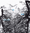
Fig. 3 Radiograph of a human necropsy specimen injected with contrast medium shows the arterial vascularization of the ventricular septum. The ventricular free walls have been removed, and only the anterior (right-sided) and posterior descending (left-sided) arteries and septal branches are shown. Note the rich collateral network, which connects the neighboring septal branches. All the collateral vessels are 0.1 to 0.3 mm in diameter. (Reproduced, with permission, from: Fulton WFM. The coronary arteries; arteriography, microanatomy, and pathogenesis of obliterative coronary artery disease. Springfield [IL]: Charles C. Thomas; 1965. p. 83–8.41) * = AV node artery; ** = 1st septal branch, primary target of theoretical septal ablation; LAD = left anterior descending artery; RCA−PDA = right coronary artery−posterior descending artery
None
Fig. 7 A) Preoperative left coronary angiogram in the right anterior oblique projection. Four branches (1–4) arising from the proximal left anterior descending coronary artery are potential candidates for alcohol infusion. On echocardiography, selective injection of contrast material into the first 2 branches showed staining of only the upper septal portions, above systolic anterior motion (SAM). B) The 3rd branch is an epicardial right ventricular branch, and the 4th branch supplies the septum at the level of SAM. C) A subselective angiogram of the 4th septal branch, which was eventually treated with alcohol septal ablation.
None
Fig. 4 Echocardiographic images obtained by injecting the 1st (A) and 2nd (B) septal branches. Note that the injection of albumex at the 1st septal branch reveals the uppermost portion of the septum (arrow), above the level of systolic anterior motion (SAM) (in A). In B, the contrast is shown (arrow) at the level of SAM. (C) Diagram of views A and B identifies the target areas in the 1st (A) and 2nd (B) septal branches. The 2nd septal branch is the ideal target for alcohol septal ablation (at the level of SAM of the mitral valve [MV]).
None
Fig. 5 Preoperative coronary angiograms from a patient with a complex coronary anomaly involving right-sided origination of all 3 major coronary vessels. A) The right coronary artery originates normally. B) An anterior artery (with a prepulmonary course) leads to the proximal section of the left anterior descending artery and its mid-septal branches. View B also shows the ectopic origin of the two 1st septal branches (asterisks), which course intraseptally and were treated successfully with alcohol septal ablation. C) A posterior, retroaortic vessel leads to the ramus, diagonal, and circumflex arteries.
None
Fig. 6 A) Right anterior oblique angiogram of the left coronary artery (LCA) shows 2 parallel septal branches (right-sided branch, R; left-sided branch, L). B) Left anterior oblique cranial angiogram of the LCA shows the sidedness of the 2 septal branches. The left branch (L) was treated, but the right branch (R) supplied only the right side of the ventricular septum and was not treated.
None
Fig. 8 A) Angiogram in the right anterior oblique projection shows the 1st septal branch (asterisk), which is the target of alcohol septal ablation. B) Angiogram of the right coronary artery in the left anterior oblique view reveals the subselective location of a 0.014-inch guidewire at the atrioventricular (AV) node artery. C) Subselective injection of a contrast agent at the AV node artery (asterisk). Infusion of acetylcholine (20 μg) at this level induced transient AV block (5-sec asystole, while temporary pacing was withheld). (See text.)
None
Fig. 9 Postoperative cardiac magnetic resonance images, all from the same patient, in the longitudinal (4-chamber) (A, B) and cross-sectional (C, D) views. Comparisons of the early images, obtained on day 2 (A, C) and the late images, obtained on day 90 (B, D) clearly show that alcohol septal ablation caused negative remodeling of the scar tissue (length of scar is indicated by double-headed arrows), which led to reduction of the subaortic gradient from 100 to 0–15 mmHg. (Images courtesy of Y.C. Cheong, MD, MRCP, Department of Radiology, St. Luke's Episcopal Hospital, Houston, Texas)

References

    1. Sigwart U. Non-surgical myocardial reduction for hypertrophic obstructive cardiomyopathy. Lancet 1995;346:211–4. - PubMed
    1. Shamim W, Yousufuddin M, Wang D, Henein M, Seggewiss H, Flather M, et al. Nonsurgical reduction of the interventricular septum in patients with hypertrophic cardiomyopathy. N Engl J Med 2002;347:1326–33. [Retraction in: Coats AJ, Henein M, Flather M, Sigwart U, Seggewiss H, Wang D, et al. N Engl J Med 2003;348:951.] - PubMed
    1. Knight C, Kurbaan AS, Seggewiss H, Henein M, Gunning M, Harrington D, et al. Nonsurgical septal reduction for hypertrophic obstructive cardiomyopathy: outcome in the first series of patients. Circulation 1997;95:2075–81. - PubMed
    1. Seggewiss H, Gleichmann U, Faber L, Fassbender D, Schmidt HK, Strick S. Percutaneous transluminal septal myocardial ablation in hypertrophic obstructive cardiomyopathy: acute results and 3-month follow-up in 25 patients. J Am Coll Cardiol 1998;31:252–8. - PubMed
    1. Seggewiss H, Faber L, Gleichmann U. Percutaneous transluminal septal ablation in hypertrophic obstructive cardiomyopathy. Thorac Cardiovasc Surg 1999;47:94–100. - PubMed

MeSH terms