

Review
doi: 10.1002/dvdy.21335.
Caenorhabditis elegans germ line: a model for stem cell biology
Affiliations
- PMID: 17948315
- PMCID: PMC2949268
- DOI: 10.1002/dvdy.21335
Item in Clipboard
Review
Caenorhabditis elegans germ line: a model for stem cell biology
Dev Dyn.
2007 Dec.
Abstract
Like many stem cell systems, the Caenorhabditis elegans germ line contains a self-renewing germ cell population that is maintained by a niche. Although the exact cellular mechanism for self-renewal is not yet known, three recent studies shed considerable light on the cell cycle behavior of germ cells, including a support for significant and plastic renewal potential. This review brings together the results of the three recent cell-based studies, places them in the context of previous work, and discusses future perspectives for the field.
2007 Wiley-Liss, Inc
Figures

The C. elegans hermaphrodite adult germline proliferative zone. Top: cartoon depicting organization of the gonad and germ line within the worm. Middle: DAPI-stained distal end of a dissected gonad, indicating features that distinguish proliferative from transition zones. Modified (with permission) from Maciejowski et al. (2006). Bottom: proposed sub-zones within the proliferative zone. The transition zone is indicated by shading. Individual wild-type animals vary in the exact borders of these regions; dotted lines indicate border regions. CD: Cell-diameter distance.

The germ line and distal tip cell. A and B are A and D, respectively from Figure 7, Crittenden et al. (2006); reproduced with permission. A: Projection of two longitudinal confocal sections through the adult gonad showing the rachis and the extent of DTC intercalation with distal-most germ cells. Green, GFP; red, anti-GLP-1; blue, TO-PRO-3. B: Projection of confocal z-series. Green, GFP Images are projections of confocal z-series. Green, GFP (qIs56); pink, anti-PH3; blue, TO-PRO-3. Green triangle indicates proximal extent of extensive DTC contact; yellow circle, proximal extent of longest DTC process; diamond with dashed line indicates the proliferative zone/transition zone boundary. C: Modified from Figure 5, Hall et al. (1999) with permission. Scanning electron micrograph showing DTC body and cytoplasmic processes on the surface of germ “cells”. D and E: Transmission electron micrograph of a longitudinal section through the distal end of an L4 gonad showing the extent to which distal germ cells are bound by the DTC. N = DTC nucleus. (D. Hall, unpublished). E: The DTC has been outlined in grey and shaded in green; the borders of germ cells traced in black. Arrowhead indicates distal direction.

Diagrams of relationships of selected proposed interactions between signaling pathways that influence proliferation and differentiation. A: simple relationship between GLP-1 activity and germ cell fate. B: Additional interactions downstream of the GLP-1/Notch signaling pathway. Protein families/orthologs are indicated in gray. See text for further explanation.
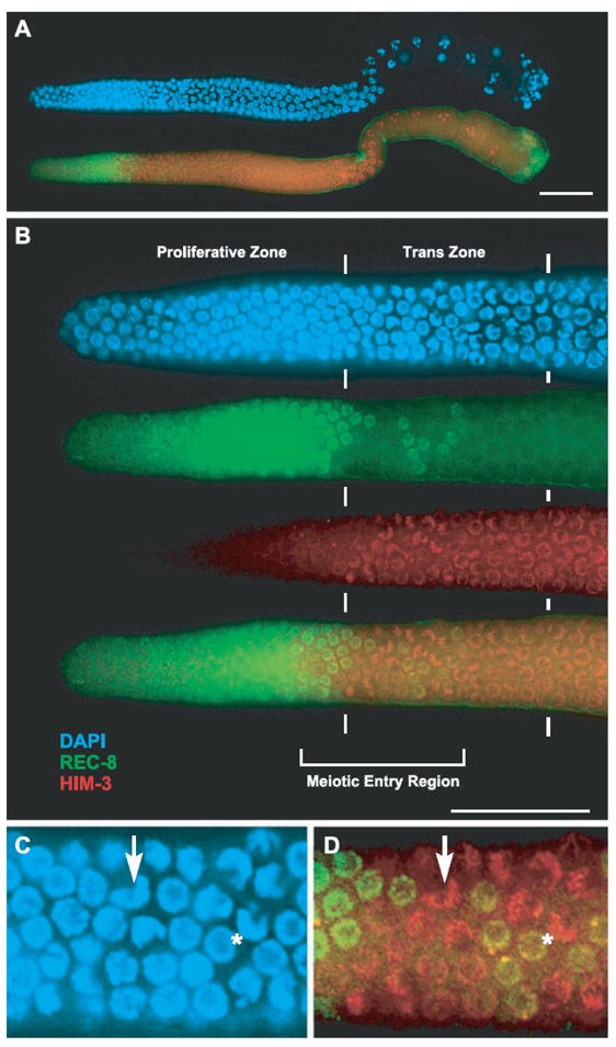
REC-8 and HIM-3 staining in a wild-type hermaphrodite. (Figure and legend reproduced with permission from Figure 3, Hansen et al., 2004a). Dissected gonad arm from an adult hermaphrodite stained with DAPI (blue), REC-8 antibodies (green), and HIM-3 antibodies (red). (A) Shows the entire gonad arm with distal to the left and proximal to the right. (B) Shows a blow-up of the distal mitotic region and the transition zone of the same gonad arm in (A); however, REC-8 and HIM-3 antibody staining are also shown separately. The boundaries of the mitotic and transition zones are demarcated with vertical lines. The location of the meiotic entry region is within the horizontal racket. (C) and (D) are further blow-ups of the same gonad arm stained with DAPI (C) and anti-REC-8 and anti-HIM-3 specific antibodies (D). The region shown is part of the meiotic entry region with the arrow pointing to a representative transition zone nucleus with crescent-shaped DAPI staining. The asterisk is beside a REC-8-positive nucleus that has a non-crescent-shaped DNA organization but is further from the DTC than the HIM-3-positive nucleus with crescent-shaped DAPI staining (arrow). This and other REC-8-positive nuclei in the meiotic entry region are more yellow than in the proliferative region, which appears to be due to faint HIM-3 staining and may represent cells that are in the transition from REC-8-positive to HIM-3-positive. Scale bars = 20 μm.

S phase, mitosis, and selected relative steady-state protein levels as a function of distance from the distal tip. A–E: charts were scaled to align axes. Exact scaling was not possible for D and E; arrows between C and D indicate positions of cell-diameter marks for D and E relative to A, B, and C. A: Chart and legend reproduced from Figure 1C with permission from Jaramillo-Lambert et al., 2007. “Chart showing the percent of Cy3-dUTP labeled nuclei at the indicated rows (nuclear diameters) from the distal tip cell. Solid line indicates the proliferative zone; dashed lines indicate the region where both proliferative and transition zone nuclei are observed. Seven gonads were examined; standard error is shown.” B and C: Charts and legend reproduced from Figure 2A and 2B with permission from Crittenden et al., 2006. “S- and M-phase indices scored in hermaphrodites 24 h past L4. Solid line indicates region of mitotic cell cycle in all germ lines. Dotted line spans positions of MR/TZ boundaries in all germ lines scored. Dashed line indicates region of meiotic prophase in all germ lines. [B] Labeling-index. % BrdU-positive cells at positions along the distal-proximal axis. Animals were fed BrdU for 15 min with no chase. Bars, 95% confidence limit. Data from 12 germ lines. [C] Mitotic index. Percent PH3 cells at positions along the distal-proximal axis. Data from 102 germ lines.” D and E: Charts and legend modified from Figure 2B and 2C, Maciejowski et al., 2006), with permission. The blue and red lines were intensified for clarity. D: “The intensity function from the large data set with 95% confidence intervals (dashed lines) for the wild-type (blue) plus the selected glp-1(ar202) mutants (red).” E. “The wild-type intensity profile from panel [D] overlaid with the approximate steady-state protein concentration levels estimated from previous publications (Crittenden et al., 1994; Hansen et al., 2004b; Jones et al., 1996; Lamont et al., 2004). For GLP-1, we depict the region of high penetrance of high-level cell membrane-associated protein only; significant levels of internal GLP-1 are present in the germ line proximal to CD 12 (see Crittenden et al., 1994, Figs. 4–6 for details). Levels are arbitrary between a maximum for each protein (indicated on graph) and a minimum (where y = 0).”
References
-
- Ambros V. Cell cycle-dependent sequencing of cell fate decisions in Caenorhabditis elegans vulva precursor cells. Development. 1999;126:1947–1956. - PubMed
-
- Austin J, Kimble J. glp-1 is required in the germ line for regulation of the decision between mitosis and meiosis in C. elegans. Cell. 1987;51:589–599. - PubMed
-
- Beanan M, Strome S. Characterization of a germ-line proliferation mutation in C. elegans. Development. 1992;116:755–766. - PubMed
-
- Berry L, Westlund B, Schedl T. Germ-line tumor formation caused by activation of glp-1, a Caenorhabditis elegans member of the Notch family of receptors. Development. 1997;124:925–936. - PubMed
-
- Brauchle M, Baumer K, Gonczy P. Differential activation of the DNA replication checkpoint contributes to asynchrony of cell division in C. elegans embryos. Curr Biol. 2003;13:819–827. - PubMed
Publication types
MeSH terms
Substances
Grants and funding
LinkOut - more resources
Full Text Sources
