. 2008 Mar;123(3):447-55.
doi: 10.1111/j.1365-2567.2007.02711.x. Epub 2007 Oct 19.

Antigen processing and CD24 expression determine antigen presentation by splenic CD4+ and CD8+ dendritic cells

Affiliations

Antigen processing and CD24 expression determine antigen presentation by splenic CD4+ and CD8+ dendritic cells

David Askew et al. Immunology. 2008 Mar.

Abstract

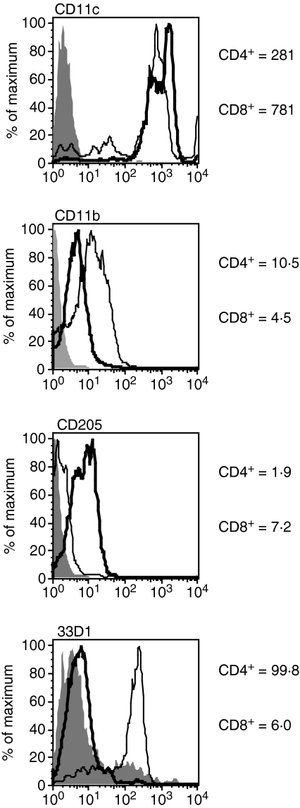
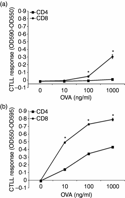
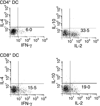
To examine heterogeneity in dendritic cell (DC) antigen presentation function, murine splenic DCs were separated into CD4+ and CD8+ populations and assessed for the ability to process and present particulate antigen to CD4+ and CD8+ T cells. CD4+ and CD8+ DCs both processed exogenous particulate antigen, but CD8+ DCs were much more efficient than CD4+ DCs for both major histocompatibility complex (MHC) class II antigen presentation and MHC class I cross-presentation. While antigen processing efficiency contributed to the superior antigen presentation function of CD8+ DCs, our studies also revealed an important contribution of CD24. CD8+ DCs were also more efficient than CD4+ DCs in inducing naïve T cells to acquire certain effector T-cell functions, for example generation of cytotoxic CD8+ T cells and interferon (IFN)-gamma-producing CD4+ T cells. In summary, CD8+ DCs are particularly potent antigen-presenting cells that express critical costimulators and efficiently process exogenous antigen for presentation by both MHC class I and II molecules.


Figures

Figure 1
Figure 1
Phenotypically distinct splenic dendritic cell (DC) populations. Freshly isolated CD4+ or CD8+ DCs were stained for expression of CD11c, CD11b, DEC-205 and 33D1 to distinguish between the two populations. CD4+ DCs expressed higher levels of CD11b and lower levels of DEC-205 when compared with CD8+ DCs. 33D1 was effective in differentiating CD4+ DCs from CD8+ DCs.
Figure 2
Figure 2
CD8+ dendritic cells (DCs) are more efficient than CD4+ DCs for processing and presentation of latex-ovalbumin (L-OVA) to T hybridoma cells. CD4+ or CD8+ DCs (2 × 10⁴) were cultured with L-OVA for 1 hr prior to addition of 105 CD8OVA1·3 [a, major histocompatibility complex class I (MHC-I)-restricted] or DOBW (b, MHC-II-restricted) T hybridoma cells. Cultures were then incubated at 37° for 24 hr, and supernatants were collected for a CTLL assay to assess interleukin (IL)-2 production. (a) CD8OVA1·3 responses. (b) DOBW responses. Data points are mean ± standard deviation (*, P < 0·01 by Student's t-test). When not visible, error bars are smaller than the symbol. Results are representative of five independent experiments.
Figure 3
Figure 3
CD8+ dendritic cells (DCs) are more efficient than CD4+ DCs for induction of naïve CD4+ and CD8+ T cell proliferative responses. CD4+ or CD8+ DCs (4 × 10⁴) were cultured with latex-ovalbumin (L-OVA) for 1 hr prior to addition of 105 T cells. Cultures were then incubated at 37° for 96 hr with 1 µCi [3H]thymidine added for the last 8 hr. Cells were harvested and [3H]thymidine incorporation was assessed. (a) Major histocompatibility complex class I (MHC-I)-restricted OT-I T-cell responses. (b) MHC-II-restricted OT-II T-cell responses. Data points represent mean ± standard deviation (*, P < 0·01 by Student's t-test). When not visible, error bars are smaller than the symbol. Results are representative of six independent experiments. CPM, counts per minute.
Figure 4
Figure 4
CD24 expression distinguishes CD4+ and CD8+ dendritic cells (DCs). Splenic CD4+ and CD8+ DCs were stained for expression of costimulatory molecules CD24, CD80 and CD86 and assessed by flow cytometry. Bold line, CD8+ DCs; thin line, CD4+ DCs. Shaded histogram, staining of CD8+ DCs with isotype-matched negative control antibody (CD4+ DCs gave similar staining with control antibody). Results are representative of five independent experiments.
Figure 5
Figure 5
CD24 functions as a costimulatory molecule for CD4+ and CD8+ T cells. CD8+ dendtritic cells (DCs) (4 × 10⁴) were cultured for 1 hr with latex-ovalbumin (L-OVA) (1 µg/ml) with or without blocking antibodies (5 µg/ml) to CD24 (clone M1/69), CD80 (clone 16·10A1), and/or CD86 (clone GL1) for 1 hr. The isotype control is represented by rat immunoglobulin G2a (IgG2a) (clone R35-95). There was no change in T-cell proliferation when other isotype controls were used (rat IgG2b or Armenian hamster IgG). T cells (105) were then added and cultures were incubated for 96 hr, with 1·0 µCi [3H]thymidine added for the last 8 hr. Cells were then harvested and [3H]thymidine incorporation was assessed. (a) Major histocompatibility complex class I (MHC-I)-restricted OT-I responses. (b) MHC-II-restricted OT-II responses. Data points represent mean ± standard deviation (*, P < 0·01 by Student's t-test). The results are representative of three independent experiments. CPM, counts per minute.
Figure 6
Figure 6
CD8+ dendritic cells (DCs) are more efficient at generating CD8+ cytotoxic T lymphocytes (CTLs). CD4+ or CD8+ DCs (2 × 10⁵) were cultured with 2 × 10⁶ OT-I CD8+ T cells in 24-well plates for 5 days. EL4 cells were incubated with [3H]thymidine and ovalbumin (OVA323-339), and labelled EL4 target cells (104 cells/well) were plated in a 96-well plate with the indicated ratio of cultured OT-I CD8+ T cells for 5 hr at 37°. The percentage cytotoxicity was determined as described in ‘Materials and methods’. Data points represent mean ± standard deviation (*, P < 0·01 by Student's t-test). When not visible, error bars are smaller than the symbol Results are representative of six independent experiments.
Figure 7
Figure 7
CD4+ and CD8+ dendritic cells (DCs) both generate T helper type 1 (Th1)-like responses by CD4+ T cells. CD4+ or CD8+ DCs (4 × 10⁴) were cultured for 1 hr with latex-ovalbumin (L-OVA) (1 µg/ml). OT-II cells were then added for 96 hr. Cells were then washed and added to 96-well plates coated with anti-CD3 in the presence of 1 µg/ml brefeldin A for 5–6 hr. Cells were washed, stained with anti-CD3, fixed with formaldehyde, labelled with anticytokine antibodies in the presence of saponin, and assessed by flow cytometry. Results are representative of four independent experiments. IFN, interferon; IL, interleukin.
Figure 8
Figure 8
CD4+ and CD8+ dendritic cells (DCs) support production of interferon (IFN)-γ and interleukin (IL)-2 by ovalbumin (OVA)-specific CD4+ T cells. Latex-ovalbumin (L-OVA)-pulsed CD4+ or CD8+ T cells were cultured with OT-II T cells for 96 hr. (a) Cell supernatant was collected and production of cytokines was determined using a cytokine antibody array. (b) The relative levels of each cytokine were compared to those for T cells cultured in media alone, as described in ‘Material and methods’. CD8+ DCs promoted increased production of IL-2 and IFN-γ when compared with T cells cultured with CD4+ DCs (*, P < 0·01 by Student's t-test). Results are representative of three independent experiments.

References

    1. Pure E, Inaba K, Crowley MT, Tardelli L, Witmer-Pack MD, Ruberti G, Fathman G, Steinman RM. Antigen processing by epidermal Langerhans cells correlates with the level of biosynthesis of major histocompatibility complex class II molecules and expression of invariant chain. J Exp Med. 1990;172:1459–69. - PMC - PubMed
    1. Reis e Sousa C, Stahl PD, Austyn JM. Phagocytosis of antigens by Langerhans cells in vitro. J Exp Med. 1993;178:509–19. - PMC - PubMed
    1. Koch F, Heufler C, Kampgen E, Schneeweiss D, Bock G, Schuler G. Tumor necrosis factor alpha maintains the viability of murine epidermal Langerhans cells in culture, but in contrast to granulocyte/macrophage colonly-stimulating factor, without inducing their functional maturation. J Exp Med. 1990;171:159–71. - PMC - PubMed
    1. Sallusto F, Cella M, Danieli C, Lanzavecchia A. Dendritic cells use macropinocytosis and the mannose receptor to concentrate macromolecules in the major histocompatibility complex class II compartment. downregulation by cytokines and bacterial products. J Exp Med. 1995;182:389–400. - PMC - PubMed
    1. Dieu M-C, Vanbervliet B, Vicari A, et al. Selective recruitment of immature and mature dendritic cells by distinct chemokines expressed in different anatomic sites. J Exp Med. 1998;188:373–86. - PMC - PubMed

Publication types

LinkOut - more resources