

Kupffer cells are central in the removal of nanoparticles from the organism
- PMID: 17949501
- PMCID: PMC2146996
- DOI: 10.1186/1743-8977-4-10
Kupffer cells are central in the removal of nanoparticles from the organism
Abstract
Background: The study aims at revealing the fate of nanoparticles administered intravenously and intraperitoneally to adult female mice, some of which were pregnant. Gold nanoparticles were chosen as a model because these particles have been found to be chemically inert and at the same time are easily traced by autometallography (AMG) at both ultrastructural and light microscopic levels.
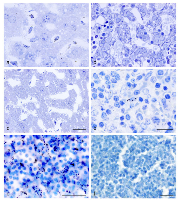
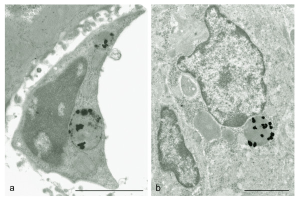
Results: Gold nanoparticles were injected intravenously (IV) or intraperitoneally (IP) and traced after 1, 4 or 24 hours. For IV injections 2 and 40 nm particles were used; for IP injections 40 nm particles only. The injected nanoparticles were found in macrophages only, and at moderate exposure primarily in the Kupffer cells in the liver. IV injections resulted in a rapid accumulation/clustering of nanoparticles in these liver macrophages, while the uptake in spleen macrophages was moderate. IP injections were followed by a delayed uptake in the liver and included a moderate uptake in macrophages located in mesenteric lymph nodes, spleen and small intestine. Ultrastructurally, the AMG silver enhanced nanocrystals were found in lysosome-like organelles of the Kupffer cells and other macrophages wherever located.Accumulations of gold nanoparticles were not found in any other organs analysed, i.e. kidneys, brain, lungs, adrenals, ovaries, placenta, and fetal liver, and the control animals were all void of AMG staining.
Conclusion: Our results suggest that: (1) inert gold nanoparticles do not penetrate cell membranes by non-endocytotic mechanisms, but are rather taken up by endocytosis; (2) gold nanoparticles, independent of size, are taken up primarily by Kupffer cells in the liver and secondarily by macrophages in other places; (3) gold nanoparticles do not seem to penetrate the placenta barrier; (4) the blood-brain barrier seems to protect the central nervous system from gold nanoparticles; (5) 2 nanometer gold particles seem to be removed not only by endocytosis by macrophages, and we hypothesize that part of these tiny nanoparticles are released into the urine as a result of simple filtration in the renal glomeruli.
Figures
Similar articles
-
Biodistribution of gold nanoparticles in mouse lung following intratracheal instillation.Chem Cent J. 2009 Nov 20;3:16. doi: 10.1186/1752-153X-3-16. Chem Cent J. 2009. PMID: 19930546 Free PMC article.
-
Protracted elimination of gold nanoparticles from mouse liver.Nanomedicine. 2009 Jun;5(2):162-9. doi: 10.1016/j.nano.2008.11.002. Epub 2009 Feb 12. Nanomedicine. 2009. PMID: 19217434
-
Penetration of pegylated gold nanoparticles through rat placental barrier.Bull Exp Biol Med. 2014 Jul;157(3):383-5. doi: 10.1007/s10517-014-2572-3. Epub 2014 Jul 29. Bull Exp Biol Med. 2014. PMID: 25065320
-
How to detect gold, silver and mercury in human brain and other tissues by autometallographic silver amplification.Neuropathol Appl Neurobiol. 1994 Oct;20(5):454-67. doi: 10.1111/j.1365-2990.1994.tb00996.x. Neuropathol Appl Neurobiol. 1994. PMID: 7845531 Review.
-
Cooperation of liver cells in health and disease.Adv Anat Embryol Cell Biol. 2001;161:III-XIII, 1-151. doi: 10.1007/978-3-642-56553-3. Adv Anat Embryol Cell Biol. 2001. PMID: 11729749 Review.
Cited by
-
Prior lung inflammation impacts on body distribution of gold nanoparticles.Biomed Res Int. 2013;2013:923475. doi: 10.1155/2013/923475. Epub 2013 Jan 13. Biomed Res Int. 2013. PMID: 23509805 Free PMC article.
-
Nuclear VEGFR-2 Expression of Hepatocytes Is Involved in Hepatocyte Proliferation and Liver Regeneration During Chronic Liver Injury.In Vivo. 2021 May-Jun;35(3):1473-1483. doi: 10.21873/invivo.12400. In Vivo. 2021. PMID: 33910825 Free PMC article.
-
Some inferences from in vivo experiments with metal and metal oxide nanoparticles: the pulmonary phagocytosis response, subchronic systemic toxicity and genotoxicity, regulatory proposals, searching for bioprotectors (a self-overview).Int J Nanomedicine. 2015 Apr 16;10:3013-29. doi: 10.2147/IJN.S80843. eCollection 2015. Int J Nanomedicine. 2015. PMID: 25945048 Free PMC article. Review.
-
Treating metastatic cancer with nanotechnology.Nat Rev Cancer. 2011 Dec 23;12(1):39-50. doi: 10.1038/nrc3180. Nat Rev Cancer. 2011. PMID: 22193407 Review.
-
Development of 68Ga-Labeled Hepatitis E Virus Nanoparticles for Targeted Drug Delivery and Diagnostics with PET.Mol Pharm. 2022 Aug 1;19(8):2971-2979. doi: 10.1021/acs.molpharmaceut.2c00359. Epub 2022 Jul 20. Mol Pharm. 2022. PMID: 35857429 Free PMC article.
References
-
- Danscher G, Norgaard JO. Light microscopic visualization of colloidal gold on resin-embedded tissue. J Histochem Cytochem. 1983;31:1394–1398. - PubMed
LinkOut - more resources
Full Text Sources
Other Literature Sources
