. 2008 May;32(5):761-70.
doi: 10.1016/j.leukres.2007.08.023. Epub 2007 Oct 18.

KIT regulates tyrosine phosphorylation and nuclear localization of beta-catenin in mast cell leukemia

Affiliations

KIT regulates tyrosine phosphorylation and nuclear localization of beta-catenin in mast cell leukemia

Tomohiro Kajiguchi et al. Leuk Res. 2008 May.

Abstract

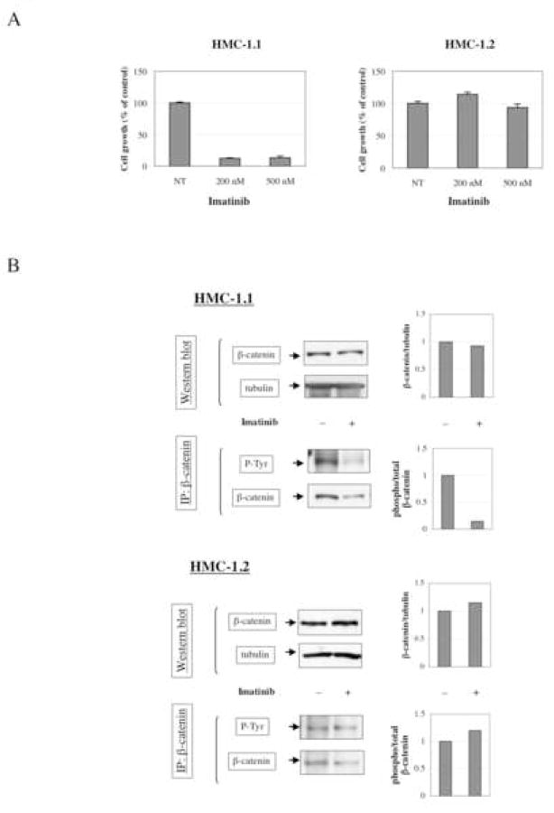
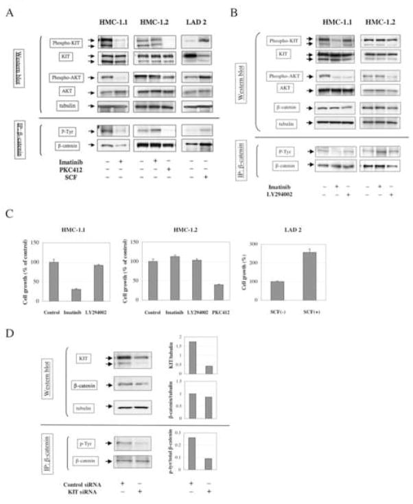
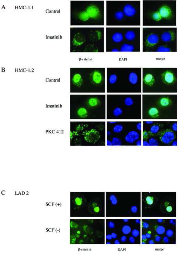
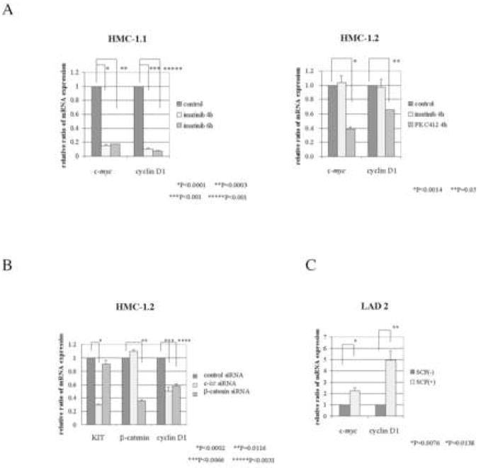
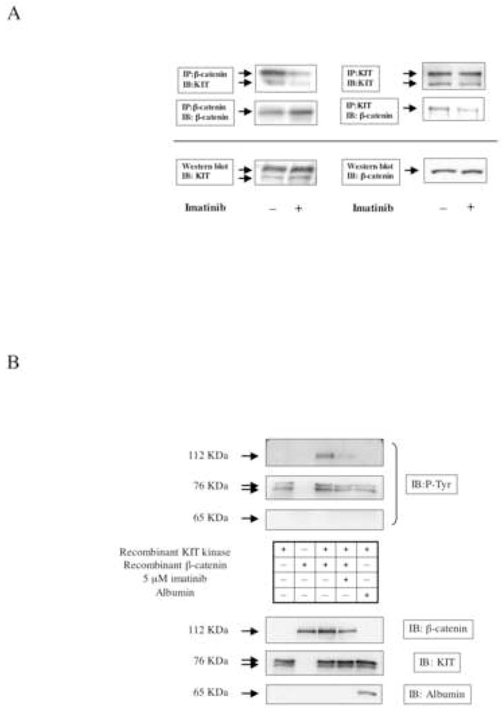
Gain-of-function mutations in the proto-oncogene c-kit that induce constitutive kinase activity of its product, KIT protein, are characteristic of human mast cell disease and are believed to play a central role in mast cell leukemia oncogenesis, proliferation and survival. Nuclear overexpression of the Wnt effector beta-catenin and deregulated beta-catenin nuclear signaling can promote malignant transformation in solid tumors and hematologic malignancies. However, a role for beta-catenin in mast cell leukemia has not been described. Nuclear accumulation of beta-catenin is upregulated by its tyrosine phosphorylation, a process that can be exacerbated by deregulated expression of oncogenic tyrosine kinases. Here, we investigated the relationship between activated KIT and beta-catenin signaling in mast cell leukemia. Beta-catenin was tyrosine-phosphorylated in cells with KIT activated by either gain-of-function mutation or incubation with the KIT ligand stem cell factor. Beta-catenin tyrosine phosphorylation depended on KIT activity but not on PI3K-AKT activation. Tyrosine phosphorylation of beta-catenin was associated with its nuclear localization and enhanced transcription of target genes c-myc and cyclin D1. Endogenous KIT and beta-catenin were found to associate in mast cell leukemia cells, and in vitro kinase assay demonstrated that active KIT phosphorylates tyrosine residues of beta-catenin directly. Aberrant beta-catenin-driven transcription caused by deregulated KIT may represent a significant new target for treatment of mast cell leukemia.


Figures

Figure 1
Figure 1
Imatinib suppresses tyrosine-phosphorylation of β-catenin in imanitib-sensitive mast cell leukemia cells. A: Effect of imatinib on cell growth of HMC-1.1 and HMC-1.2 cells. Cell growth was monitored by XTT assay as described in “Methods”. Cells were incubated with or without indicated concentration of imatinib for 24 h. Mean (+/− standard deviation) of three independent experiments are shown. B: Effect of imatinib on expression and tyrosine phosphorylation of β-catenin. To detect tyrosine phosphorylation of β-catenin, 1 mg of total protein from cell lysates was precipitated using 2 μg of anti-β-catenin antibody. Proteins were resolved by reducing 10% SDS-PAGE, electrotransferred to PVDF membrane, and western blotted using corresponding antibodies. Optical density ratio of β-catenin/tubulin and tyrosine phosphorylated/total β-catenin (phospho/total β-catenin) was measured using a GS-800 densitometer with Quantity One software.
Figure 2
Figure 2
KIT activation is related to β-catenin tyrosine phosphorylation status, but does not depend on PI3K/AKT activation. A: HMC-1.1 and HMC-1.2 cells were treated with or without the KIT inhibitors imatinib (500 nM for 3h) or PKC412 (250 nM for 3h). LAD 2 was starved of SCF for 48 h, and then treated with or without rhSCF (100 ng/ml for 3h). After the treatments, cells were lysed and lysates were subjected to western blot and immunoprecipitation. The HMC1.1 β-catenin and phosphotyrosine blots are reproduced from Fig. 1B for comparison. B: HMC-1.1 and 1.2 cells were treated with or without imatinib (500 nM for 3h) or the PI3K inhibitor LY294002 (5 μM for 3h). After the treatment, cells were lysed and subjected to western blot and immunoprecipitation assay. C: HMC-1.1 and 1.2 cells were treated with or without imatinib (500 nM), LY294002 (5 μM), or PKC412 (250 nM) for 24 h. LAD 2 cells were incubated with or without rhSCF (100 ng/ml) for 72 h, following an initial starvation period of 48 h. Cell proliferation was determined by XTT assay. Mean (+/− standard deviation) of three experiments are shown. D: HMC-1.2 cells were transfected with either 1 μM c-kit siRNA or control siRNA. Following transfection, cells were incubated for 48 h. After 48 h, cells were lysed and protein expression of KIT, β-catenin and tubulin was analyzed by western blot, and tyrosine phosphorylation of β-catenin was analyzed by immunoprecipitation assay. Optical density ratios of KIT/tubulin, β-catnein/tubulin and phospho/total β-catenin were determined using a GS-800 densitometer and Quantity One software.
Figure 3
Figure 3
Nuclear localization of β-catenin in activated c-kit cell lines. A, B: HMC-1.1 (A) and HMC-1.2 (B) cells were treated with or without imatinib (500 nM for 3 h) or PKC412 (250 ng/ml for 3 h), and then were cytocentrifuged onto glass slides. Cells were fixed in 3.7% formaldehyde in PBS and permeabilized with 0.2% Triton X-100. β-catenin was visualized by immunofluorescence (green, left panel). The DNA-intercalating dye DAPI was used to identify cell nuclei (blue, center panel). The right panel presents a merged image to highlight the nuclear pool of β-catenin. C: Localization of β-catenin in LAD 2 cells. After incubation with or without rhSCF, LAD 2 cells were cytocentrifuged onto glass slides. Cells were fixed and stained as above. Images were collected using a 100× objective.
Figure 4
Figure 4
Effect of KIT activation on β-catenin target gene expression in MCL cell lines. A: HMC-1.1 and 1.2 were treated with or without imatinib (500 nM) or PKC412 (250 nM) for indicated time periods. Total RNA was isolated from the cells and quantitative RT-PCR was performed in duplicate. All samples were normalized to the level of 18S ribosomal RNA. The mean of two individual experiments (+/− standard deviation) are shown. B: HMC-1.2 cells were transfected with either c-kit siRNA, β-catenin siRNA or control siRNA. Following incubation for 48 h, total RNA was isolated and quantitative RT-PCR was performed. C: LAD 2 cells were starved of SCF for 48 h and then were treated with or without rhSCF (100 ng/ml) for an additional 4 h. Total RNA was isolated from the cells and quantitative RT-PCR was performed. Where shown, p values were determined by t-test using the StatView software package (SAS, Cary, NC).
Figure 5
Figure 5
Active KIT binds to β-catenin and catalyzes β-catenin tyrosine phosphorylation. A: HMC-1.1 cells were treated with or without imanitib (500 nM for 3 h). After cell lysis, 1 mg of total protein was immunoprecipitated with an antibody recognizing either β-catenin (left upper panel) or KIT (right upper panel). After SDS-PAGE and electrotransfer, blots were probed with antibodies to both β-catenin and KIT. The level of β-catenin and KIT protein in whole cell lysates is shown in the lower panel. B: Recombinant β-catenin was incubated with recombinant active KIT in an in vitro kinase reaction buffer, as described in “Materials and Methods”. To demonstrate specificity of the reaction, albumin was substituted for β-catenin. After the reaction, proteins were resolved by SDS-PAGE and immunoblotted for phospho-tyrosine (upper panels). The membrane was stripped and re-probed with anti-β-catenin, anti-KIT and anti-albumin antibody to verify amounts of proteins present in the assay.

References

    1. Yarden Y, Kuang WJ, Yang-Feng T, Coussens L, Munemitsu S, Dull TJ, et al. Human proto-oncogene c-kit: a new cell surface receptor tyrosine kinase for an unidentified ligand. Embo J. 1987;6:3341–51. - PMC - PubMed
    1. Anderson DM, Lyman SD, Baird A, Wignall JM, Eisenman J, Rauch C, et al. Molecular cloning of mast cell growth factor, a hematopoietin that is active in both membrane bound and soluble forms. Cell. 1990;63:235–43. - PubMed
    1. Martin FH, Suggs SV, Langley KE, Lu HS, Ting J, Okino KH, et al. Primary structure and functional expression of rat and human stem cell factor DNAs. Cell. 1990;63:203–11. - PubMed
    1. Miettinen M, Lasota J. KIT (CD117): a review on expression in normal and neoplastic tissues, and mutations and their clinicopathologic correlation. Appl Immunohistochem Mol Morphol. 2005;13:205–20. - PubMed
    1. Blechman JM, Lev S, Givol D, Yarden Y. Structure-function analyses of the kit receptor for the steel factor. Stem Cells. 1993;11 (Suppl 2):12–21. - PubMed

Publication types