

Autoreactive human T-cell receptor initiates insulitis and impaired glucose tolerance in HLA DR4 transgenic mice
- PMID: 17949947
- PMCID: PMC2440666
- DOI: 10.1016/j.jaut.2007.08.001
Autoreactive human T-cell receptor initiates insulitis and impaired glucose tolerance in HLA DR4 transgenic mice
Abstract
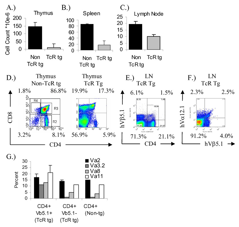
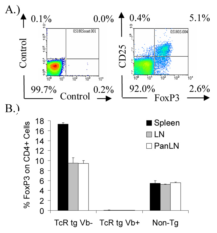
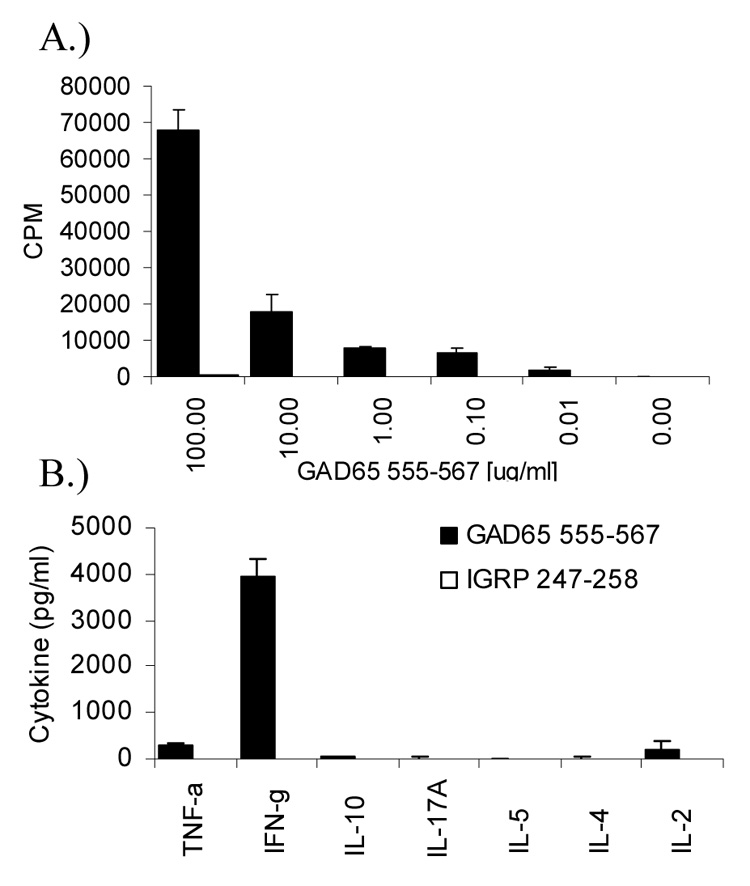
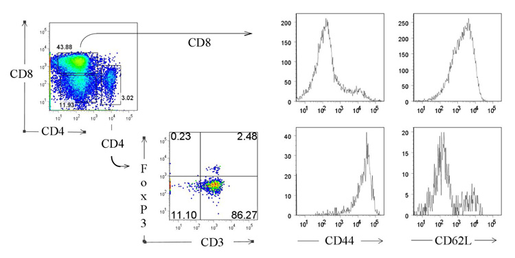
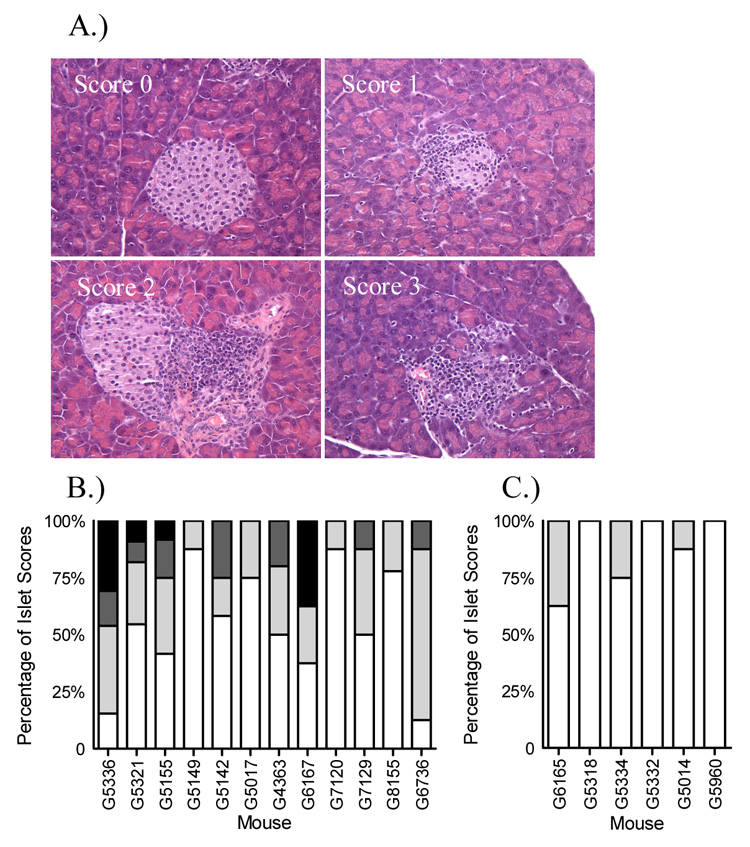
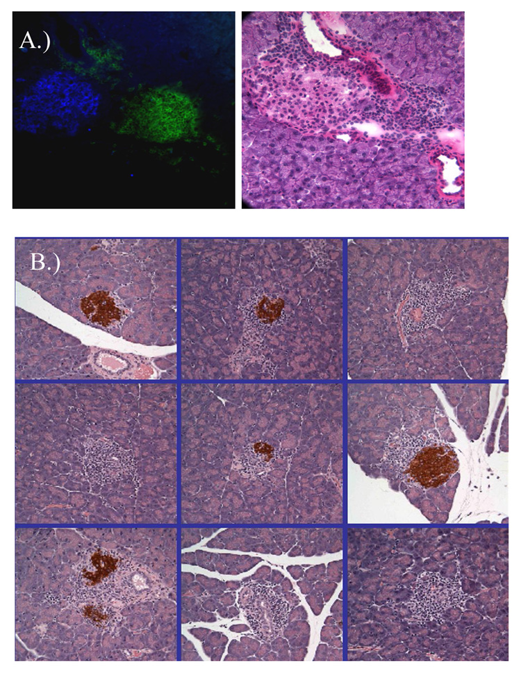
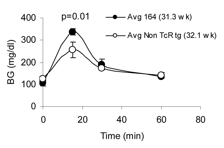
A human T-cell receptor (TcR) derived from an autoreactive T-cell specific for GAD65, from a subject at high risk for autoimmune diabetes, was introduced into HLA-DR4 transgenic mice. The source of TcR was a CD4(+) T(H)1(+) T-cell clone which responded to an immunodominant epitope of the human islet protein GAD65, an epitope shared with both GAD65 and GAD67 in the mouse. The resulting HLA-DR4/GAD-TcR transgenic mice on a Rag2(o/o)/I-Ab(o/o)/B6 background exhibited a CD4(+) infiltrate into pancreatic islets that correlated with a loss of insulin in infiltrated islets. These mice also exhibited a subclinical impaired tolerance to exogenously fed glucose as assayed by an intraperitoneal glucose tolerance test. T cells containing the GAD65/67 (555-567) responsive TcR undergo strong negative selection as evidenced by a 10-fold lower thymocyte cellularity compared to non-TcR transgenic mice, and clonotype peripheral T cells represented approximately 1% of CD4(+) T cells in Rag2 sufficient mice. Upon in vitro stimulation, GAD65/67 555-567 responsive T cells secrete interferon-gamma, minimal interleukin (IL)-2 and tumor necrosis factor-alpha, and no IL-4, IL-5, IL-10, or IL-17, consistent with a T(H)1 profile. These data demonstrate that CD4(+) T cells specific for a naturally processed epitope within GAD can specifically home to pancreatic islets and lead to impaired islet beta-cell function in diabetes-associated HLA-DR4 transgenic mice on the relatively non-autoimmune C57BL/6 background. The relatively slow progression and patchy insulitis are reminiscent of the chronic pre-clinical phase similar to a majority of human at-risk subjects, and models these indolent features of human T1D.
Figures
Similar articles
-
Antigen-specific immunomodulation for type 1 diabetes by novel recombinant antibodies directed against diabetes-associates auto-reactive T cell epitope.J Autoimmun. 2013 Dec;47:83-93. doi: 10.1016/j.jaut.2013.08.009. Epub 2013 Oct 3. J Autoimmun. 2013. PMID: 24090977
-
Dendritic Cells Guide Islet Autoimmunity through a Restricted and Uniquely Processed Peptidome Presented by High-Risk HLA-DR.J Immunol. 2016 Apr 15;196(8):3253-63. doi: 10.4049/jimmunol.1501282. Epub 2016 Mar 4. J Immunol. 2016. PMID: 26944932
-
A low antigen dose selectively promotes expansion of high-avidity autoreactive T cells with distinct phenotypic characteristics: a study of human autoreactive CD4+T cells specific for GAD65.Autoimmunity. 2010 Dec;43(8):573-82. doi: 10.3109/08916930903540424. Epub 2010 Apr 7. Autoimmunity. 2010. PMID: 20370569
-
Autoimmune diabetes: the role of T cells, MHC molecules and autoantigens.Autoimmunity. 1998;27(3):159-77. doi: 10.3109/08916939809003864. Autoimmunity. 1998. PMID: 9609134 Review.
-
HLA class II transgenic mice: models of the human CD4+ T-cell immune response.Immunol Rev. 1999 Dec;172:335-43. doi: 10.1111/j.1600-065x.1999.tb01377.x. Immunol Rev. 1999. PMID: 10631958 Review.
Cited by
-
A functional framework for interpretation of genetic associations in T1D.Curr Opin Immunol. 2012 Oct;24(5):516-21. doi: 10.1016/j.coi.2012.07.003. Epub 2012 Jul 26. Curr Opin Immunol. 2012. PMID: 22841349 Free PMC article. Review.
-
Phenotypical and functional alterations of CD8 regulatory T cells in primary biliary cirrhosis.J Autoimmun. 2010 Nov;35(3):176-80. doi: 10.1016/j.jaut.2010.06.004. Epub 2010 Jul 16. J Autoimmun. 2010. PMID: 20638239 Free PMC article.
-
Preclinical Models to Evaluate the Human Response to Autoantigen and Antigen-Specific Immunotherapy in Human Type 1 Diabetes.Front Endocrinol (Lausanne). 2022 Apr 13;13:883000. doi: 10.3389/fendo.2022.883000. eCollection 2022. Front Endocrinol (Lausanne). 2022. PMID: 35498419 Free PMC article. Review.
-
T cell recognition of autoantigens in human type 1 diabetes: clinical perspectives.Clin Dev Immunol. 2011;2011:513210. doi: 10.1155/2011/513210. Epub 2011 Jul 19. Clin Dev Immunol. 2011. PMID: 21785617 Free PMC article. Review.
-
Genetically modified human CD4(+) T cells can be evaluated in vivo without lethal graft-versus-host disease.Immunology. 2016 Aug;148(4):339-51. doi: 10.1111/imm.12613. Immunology. 2016. PMID: 27124592 Free PMC article.
References
-
- Abraham RS, Wilson SB, de SN, Jr, Strominger JL, Munn SR, David CS. NOD background genes influence T cell responses to GAD 65 in HLA-DQ8 transgenic mice. Hum. Immunol. 1999;60:583–590. - PubMed
-
- Kruisbeek Ada M. Isolation and Fractionation of Mononuclear Cell Populations. In: Coligan JE, Kruisbeek AM, Margulies DH, Shevach EM, Strober W, editors. Current Protocols in Immunology. John Wiley & Sons, Inc.; 2000. pp. 3.1.1–3.1.5.
-
- Arden B, Clark SP, Kabelitz D, Mak TW. Human T-cell receptor variable gene segment families. Immunogenetics. 1995;42:455–500. - PubMed
-
- Arnold PY, Burton AR, Vignali DA. Diabetes incidence is unaltered in glutamate decarboxylase 65-specific TCR retrogenic nonobese diabetic mice: generation by retroviral-mediated stem cell gene transfer. J. Immunol. 2004;173:3103–3111. - PubMed
-
- Bingley PJ, Bonifacio E, Williams AJ, Genovese S, Bottazzo GF, Gale EA. Prediction of IDDM in the general population: strategies based on combinations of autoantibody markers. Diabetes. 1997;46:1701–1710. - PubMed
Publication types
MeSH terms
Substances
Grants and funding
LinkOut - more resources
Full Text Sources
Other Literature Sources
Medical
Research Materials
