Review
. 2008 Apr;66(1):65-74.
doi: 10.1016/j.critrevonc.2007.09.001. Epub 2007 Oct 18.

Impact of aging on the biology of breast cancer

Affiliations
Review

Impact of aging on the biology of breast cancer

Christopher C Benz. Crit Rev Oncol Hematol. 2008 Apr.

Abstract

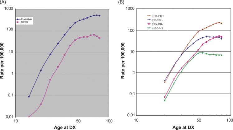
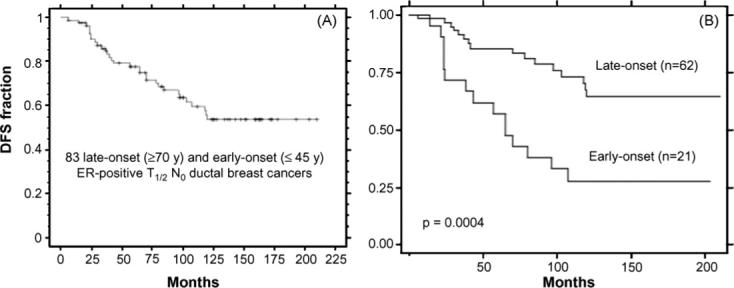
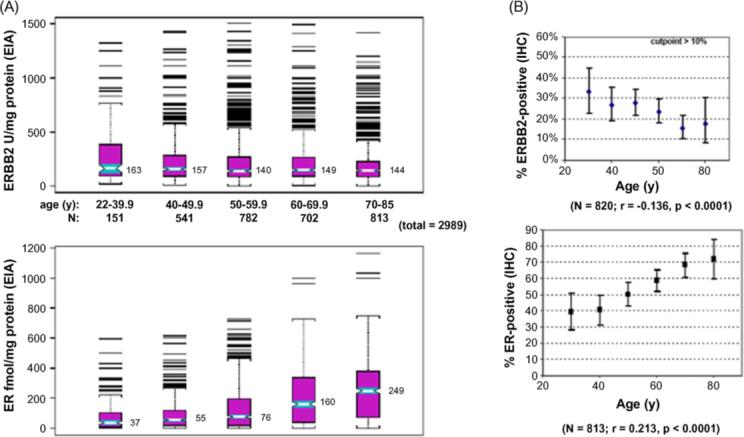
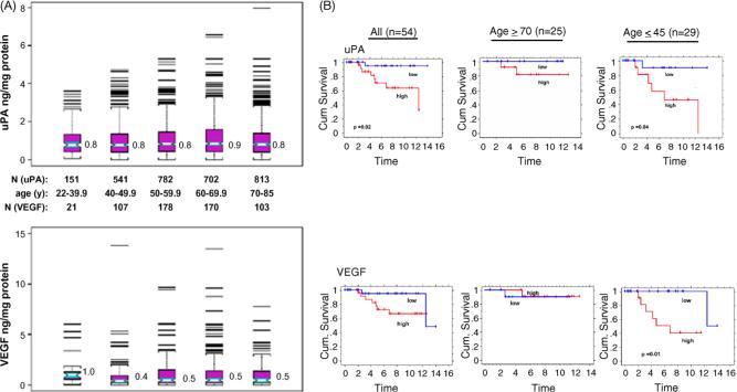
Breast cancer is a heterogeneous malignancy; its age-specific incidence profile rises exponentially until menopause and increases more slowly thereafter, reflecting the superimposition of early-onset and late-onset breast cancer rates. While early-onset breast cancers largely represent inherited or early life transforming effects on immature mammary epithelium, late-onset breast cancers likely follow extended exposures to promoting stimuli of susceptible epithelium that has failed to age normally. Among stimuli thought to promote late-onset breast tumorigenesis are the altered extracellular matrix and secreted products of senescent fibroblasts; however, the extent to which these senescent influences exist within the aging breast remains unknown. Clinical observations and biomarker studies indicate that late-onset breast cancers grow more slowly and are biologically less aggressive than early-onset breast cancers, even when controlled for hormone receptor (e.g. estrogen receptor, ER) and growth factor receptor (e.g. HER2) expression, supporting the conclusion that the biology of breast cancer is age-dependent.


Figures

Fig. 1
Fig. 1
Age-specific incidence curves (log–log plots) for overall invasive and ductal carcinoma in situ (DCIS) newly diagnosed breast cancers (panel A), and for invasive breast cancer subsets according to ER and PR status (panel B). SEER incident rates (per 100,000, including all ethnic groups) for 5-year age groups determined from the SEER reporting interval 1992−1997. Known ER and PR status was available for over 80,000 cases across all age groups; these cases consisted of 62% ER-positive/PR-positive, 13% ER-positive/PR-negative, 4% ER-negative/PR-negative, and 21% ER-negative/PR-negative. Data and figure revised from previous publication [4].
Fig. 2
Fig. 2
Kaplan–Meier disease-free survival (DFS) curve for combined set of 83 ER-positive node-negative ductal breast cancer cases untreated with adjuvant therapy (panel A), and DFS curves for late-onset (n = 62) and early-onset (n = 21) subsets (panel B). As described in the text, the selected age cohorts were well matched for numerous tumor characteristics and biomarkers and differed only by mean tumor proliferation index and high tumor grade. The significant difference in DFS outcomes shown (p = 0.0004) could not be eliminated by adjusting for subset differences in tumor grade and proliferation index. Primary data provided by A. Thor and analyzed by D. Moore.
Fig. 3
Fig. 3
Age associations for ERBB2 and ER content (panel A) or their percent overexpression (panel B) for unselected primary breast cancers from two different archives. Cryobanked Swiss tumor extracts (n = 2989) were analyzed by quantitative enzyme immunoassays (EIA), while formalin-fixed paraffin-embedded American/MGH samples (n > 800) were analyzed by immunohistochemistry and scored for percent positive staining tumor cells. Notch-boxplots show median values for each age group. Proportion plots show median% values (±95% confidence intervals) for each age group, with linear regression fit (r, Pearson's correlation coefficient) and statistical significance (p values) indicated below. Figures modified from previous publication [36].
Fig. 4
Fig. 4
Age associations for uPA and VEGF protein content from Swiss breast cancer archive (panel A), and Kaplan–Meier relapse-free survival curves based on level of breast cancer uPA and VEGF transcript expression (high, low) from American/UCSF breast cancer archive (panel B). Unselected Swiss samples were assayed as described in Fig. 3 (panel A), with figures modified from previous publication [36]. American/UCSF archive contained 54 node-negative, ER-positive breast cancer cases selected according to late-onset (≥70 years, n = 25) or early-onset (≤45 years, n = 29). Dichotomization for uPA and VEGF expression levels (high, low) was based on mean-centered transcript values, measured as previously described; figures were modified from previous publication [37]. Significant differences between the cumulative survival curves were determined by Log Rank analyses (only p values <0.05 shown).

Similar articles

Cited by

References

    1. Thun MJ, Jemal A. Cancer epidemiology. In: Kufe DW, Bast RC, Hait WN, Hong WK, Pollock RE, Weichselbaum RR, Holland JF, Frei E III, editors. Cancer 7 medicine. BC Decker Inc.; Hamilton: 2006. pp. 339–53.
    1. Jemal A, Siegel R, Ward E, Murray T, Xu J, Thun MJ. Cancer statistics 2007. CA Cancer J Clin. 2007;57:43–66. - PubMed
    1. DevCan: Probability of developing or dying of cancer software. V6.1.0. 2006. Statistical Research and Applications Branch, National Cancer Institute ( http://www.srab.cancer.gov/devcan)
    1. Quong J, Eppenberger-Castori S, Moore D, III, et al. Age-dependent changes in breast cancer hormone receptors and oxidant stress markers. Breast Cancer Res Treat. 2002;76:221–36. - PubMed
    1. Anderson WF, Pfeiffer RM, Dores GM, Sherman ME. Comparison of age distribution patterns for different histopathologic types of breast cancer. Cancer Epidemiol Biomarkers Prev. 2006;15:1899–905. - PubMed

Publication types

Substances