. 2008 Jan;37(1):96-109.
doi: 10.1016/j.mcn.2007.08.017. Epub 2007 Sep 14.

Role of astrocytes and chemokine systems in acute TNFalpha induced demyelinating syndrome: CCR2-dependent signals promote astrocyte activation and survival via NF-kappaB and Akt

Affiliations

Role of astrocytes and chemokine systems in acute TNFalpha induced demyelinating syndrome: CCR2-dependent signals promote astrocyte activation and survival via NF-kappaB and Akt

Marlon P Quinones et al. Mol Cell Neurosci. 2008 Jan.

Abstract

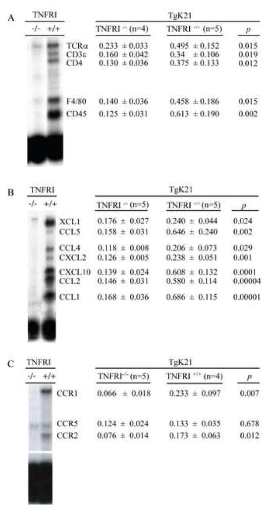
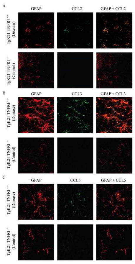
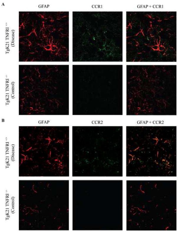
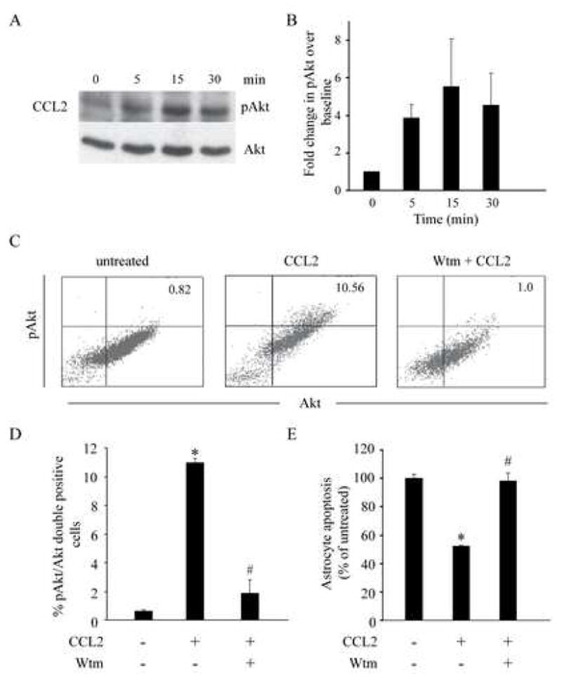
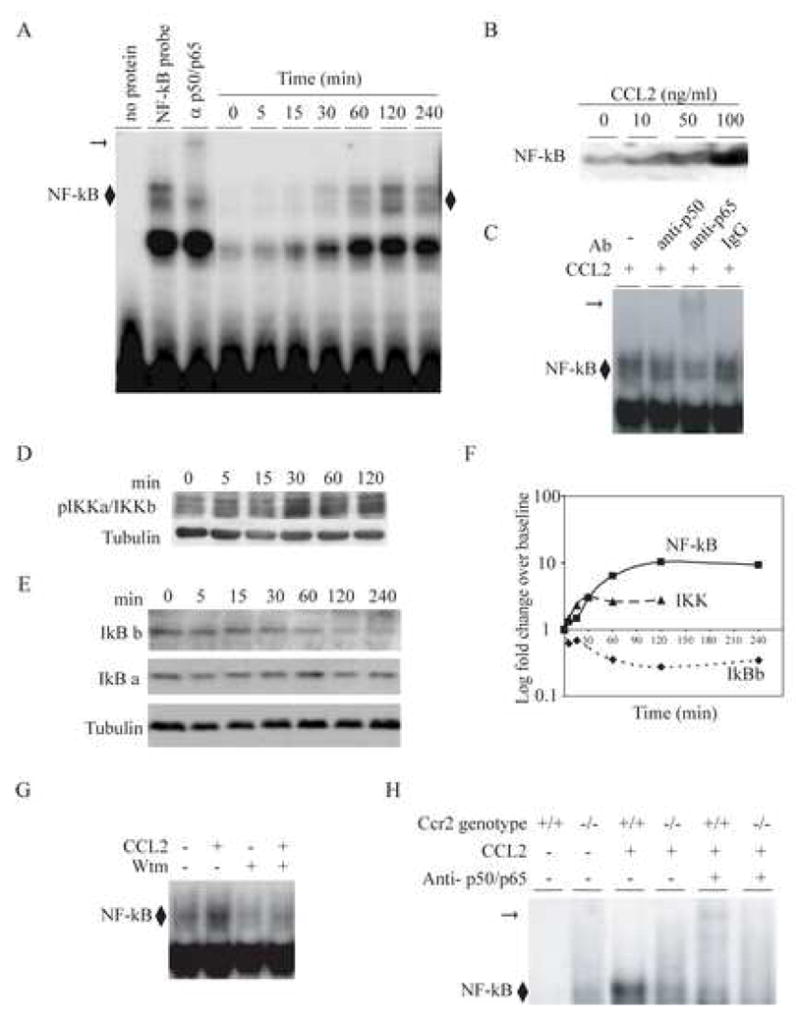
Chemotactic factors known as chemokines play an important role in the pathogenesis of multiple sclerosis (MS). Transgenic expression of TNFalpha in the central nervous system (CNS) leads to the development of a demyelinating phenotype (TNFalpha-induced demyelination; TID) that is highly reminiscent of MS. Little is known about the role of chemokines in TID but insights derived from studying this model might extend our current understanding of MS pathogenesis and complement data derived from the classic autoimmune encephalomyelitis (EAE) model system. Here we show that in TID, chemokines and their receptors were significantly increased during the acute phases of disease. Notably, the CCL2 (MCP-1)-CCR2 axis and the closely related ligand-receptor pair CCR1-CCL3 (MIP-1alpha) were among the most up-regulated during disease. On the other hand, receptors like CCR3 and CCR4 were not elevated. This significant increase in the levels of chemokines/receptors correlated with robust immune infiltration of the CNS by inflammatory cells, i.e., macrophages, and immune cells particularly T and B cells. Immunostaining and confocal microscopy, along with in vitro studies revealed that astrocytes were a major source of locally produced chemokines and expressed functional chemokine receptors such as CCR2. Using an in vitro system we demonstrate that expression of CCR2 was functional in astrocytes and that signaling via this receptor lead to activation of NF-kB and Akt and was associated with increased astrocyte survival. Collectively, our data suggests that transgenic murine models of MS are useful to dissect mechanisms of disease and that in these models, up-regulation of chemokines and their receptors may be key determinants in TID.


Figures

Figure 1
Figure 1. Demyelination and inflammatory cell infiltration of the brain in TgK21 mice
(A–B) representative staining of tissue sections of the brains of control (TgK21 TNFRI−/−) and neurologically compromised mice (TgK21 TNFRI+/+). (A–B) Brain sections cut at the level of cerebellum were stained with luxol fast blue that stains myelin light blue. Note severe demyelination, evident by lack of bluish green stain for myelin in B. (C–D) Sections derived from different brain regions were stained with H&E to better identify infiltrating cells. Note marked infiltration of mononuclear cells in the brain tissue of TgK21 TNFRI+/+ (D) mice compared with TgK21 TNFRI−/− (C). Representative sections out of many collected and analyzed in a blinded fashion by the pathologist are shown.
Figure 2
Figure 2
Infiltrating leukocytes, chemokines and chemokine receptors in the brains of K21 TNFα transgenic mice. mRNA transcripts for (A) cell type specific molecules, (B) chemokines and (C) chemokine receptors were measured in total RNA obtained from the whole brain of mice using RNA protection assay. The data are presented as the ratio of the densitometric signals of the mRNA for the gene of interest and that of a housekeeping gene (GAPDH) and. is shown as Mean ± SD.
Figure 3
Figure 3
Confocal microscope image of the brains of TgK21 mice immunolabeled with antibodies against the astrocyte marker GFAP and CCL2 (A), CCL3 (B) and CCL5 (C). Note marked increase in GFAP immunoreactivity in TNFαR+/+ mice that were commonly clinically compromised. Coronal brain sections were at the middle portion of the parietal lobes. Photomicrographs are representative of two separate experiments with each experiment having 3 mice per group.
Figure 4
Figure 4
Confocal microscope image of the brains of TgK21 mice immunolabeled with antibodies against the astrocyte marker GFAP and CCR1 (A) or CCR2 (B). Coronal brain sections were at the middle portion of the parietal lobes. Photomicrographs are representative of two separate experiments with each experiment having 3 mice per group.
Figure 5
Figure 5
CCR2 is expressed on murine astrocytes and evidence for an additional receptor for CCL2. (A), CCR2 expression on GFAP positive murine astrocytes was determined by flow cytometric analysis using monoclonal anti-mouse CCR2 and GFAP antibodies. The plot for Ccr2+/+ cells (black) is shifted to the right relative to the plot for Ccr2−/− cells (grey) and the isotype control (dashed), indicating expression of CCR2 on wild type cells but not on those lacking expression of CCR2. (B), Astrocyte migration in response to CCL2. As a functional readout for CCL2 effects, we determined the migration of Ccr2+/+ and Ccr2−/− astrocytes in response to this chemokine. Astrocyte chemotaxis assays were performed using transwells in the absence or presence of CCL2 for 20 hours. *P < 0.01 for comparison of chemotaxis of Ccr2+/+ astrocytes in the presence and absence of CCL2. #P <0.01 for comparison of chemotaxis of Ccr2+/+ and Ccr2−/− astrocytes in the presence of CCL2. **P < 0.01 for comparison of chemotaxis of Ccr2−/− astrocytes in the presence or absence of CCL2. Data are represented as the chemotaxis index (mean ± SD) derived from three independent experiments conducted in triplicate. (C), CCL2 did not alter astrocyte proliferation. Astrocytes were stimulated with CCL2 (100ng/ml) for 72 hours. During the last 16 hours of culture, cells were pulsed with [3H] thymidine. Thymidine incorporation into the DNA was detected using scintillation scanning. Data is representative of two independent experiments conducted in triplicate (mean ± SEM). (D), CCL2 decreased the spontaneous apoptosis of astrocytes in culture in a dose dependent manner. CCL2 was added to astrocytes maintained in cell culture at the indicated doses, and apoptosis was determined by staining cells with propidium iodide (PI) and Annexin V-FITC. Histograms indicate the PI-negative, Annexin V positive cells. Note, plots for cells treated with CCL2 are shifted to the left relative to plots indicating cells that were untreated. (E). CCL2 (100 ng/ml) did not decrease the spontaneous apoptosis of Ccr2−/− astrocytes, but did so in Ccr2+/+ astrocytes (mean ± SEM). The methods used were similar to those described in panel A.
Figure 6
Figure 6
Pro-survival effect of CCL2 in astrocytes was dependent on a PI 3-kinase/Akt pathway. Astrocytes were stimulated with CCL2 (100ng/ml) for the indicated time intervals and cell extracts were then prepared for the analyses. (A). CCL2-induced phosphorylation of Akt in astrocytes (upper panel). Phosphorylation of Akt was detected by western blot using an antibody against phospho-Akt (p-Akt). An antibody against Akt was used to demonstrate equal loading of the samples (lower panel). (B), summary of the kinetics of Akt activation (mean fold change ± SEM) derived from 2 independent experiments. Data are shown as fold increase in Akt phosphorylation relative to untreated cells (time = 0). (C), CCL2-induced phosphorylation of Akt was blocked by wortmannin (Wtm), a PI 3-kinase inhibitor. Astrocytes were treated with wortmannin (250nM) for 1 hour followed by stimulation with CCL2 for 15 minutes. Phosphorylation of Akt was detected using flow cytometry by dual labeling of astrocytes with antibodies against Akt and pAkt. Cells in the upper right quadrant are double positive for Akt and pAkt, indicating that they are astrocytes with phosphorylated Akt. (D), summary data (mean percent change ± SEM) for inhibition of CCL2-induced phosphorylation of Akt by wortmannin from two independent experiments conducted in triplicate. (E), Inhibition of PI 3-kinase reduced the anti-apoptotic effects of CCL2. Apoptosis of astrocytes was detected using double labeling of cells with PI and Annexin V. Data are shown as percentage (%) change (± SEM) in apoptosis relative to untreated cells (100%). For D and E, * P < 0.05 for comparison between cells stimulated vs unstimulated with CCL2, # P < 0.05 for comparison between cells treated with CCL2 vs those treated with wortmannin and CCL2.
Figure 7
Figure 7
CCL2 induced activation of the NF-κB pathway in primary murine astrocytes. Astrocytes were derived from Ccr2+/+ animals. (A), Time-dependent induction of NF-κB by CCL2 (100ng/ml). Astrocytes were treated with CCL2 for the indicated time-points and nuclear extracts were prepared for EMSAs. EMSAs were conducted using a consensus NF-κB probe and supershift assays were performed using a combination of anti-p50 and -p65 antibodies (third lane). Note, supershift in the third lane indicated by the horizontal arrow. ◆, represents NF-κB bound to the probe. Maximal nuclear translocation of NF-κB was detected at 120 min, and this time interval was therefore used in the experiments shown in panels B and C. (B), CCL2 induced nuclear translocation of NF-κB in a dose-dependent manner. Primary murine astrocytes were treated with the indicated concentrations of CCL2 for 120 min and nuclear translocation of NF-κB was detected by EMSAs. (C), CCL2 induced predominantly the translocation of the p65 component of the NF-κB complex. A supershift was detected with an anti-p65 Ab, but not anti-p50 or isototype (IgG) control Ab. (D), CCL2 activated IKKα/IKKβ in astrocytes. Astrocytes were treated with CCL2 (100ng/ml) for the indicated time intervals. Cytoplasmic extracts were prepared and probed with the indicated Abs using Western blots. Anti-tubulin antibody was used to determine equal protein loading (lower panel). Maximal phosphorylation of IKKα/IKKβ was detected at 30 min. (E), CCL2-induced NF-κB translocation is associated with degradation of IκBβ but not IκBα. Cytoplasmic extracts were prepared from astrocytes treated with CCL2 for the time intervals shown and then probed with the indicated antibodies using Western blots. (F), Summary of time kinetics of signaling events that lead to CCL2 induced NF-κB nuclear translocation. Fold differences shown in panels A to E were log transformed and plotted against time. The CCL2-induced NF-κB nuclear translocation in astrocytes was dependent on PI 3 -kinase and expression of CCR2. (G), CCL2-induced nuclear translocation of NF-κB is inhibited by wortmannin (Wtm). Astrocytes were treated with 250nM wortmannin for 1 hour before stimulation with CCL2 for 2 hours. Nuclear extracts were prepared and used for EMSA. (H), CCL2-induced nuclear translocation of NF-κB was markedly diminished in Ccr2−/− astrocytes. Nuclear extracts were derived from Ccr2+/+ and Ccr2−/− astrocytes treated with CCL2 at 100 ng/ml for 120 min. Supershifts were performed with a combination of anti-p50 and -p65 antibodies.

Similar articles

Cited by

References

    1. Akassoglou K, Adams RA, Bauer J, Mercado P, Tseveleki V, Lassmann H, Probert L, Strickland S. Fibrin depletion decreases inflammation and delays the onset of demyelination in a tumor necrosis factor transgenic mouse model for multiple sclerosis. Proc Natl Acad Sci U S A. 2004;101:6698–6703. - PMC - PubMed
    1. Akassoglou K, Bauer J, Kassiotis G, Lassmann H, Kollias G, Probert L. Transgenic models of TNF induced demyelination. Adv Exp Med Biol. 1999;468:245–259. - PubMed
    1. Akassoglou K, Bauer J, Kassiotis G, Pasparakis M, Lassmann H, Kollias G, Probert L. Oligodendrocyte apoptosis and primary demyelination induced by local TNF/p55TNF receptor signaling in the central nervous system of transgenic mice: models for multiple sclerosis with primary oligodendrogliopathy. Am J Pathol. 1998;153:801–813. - PMC - PubMed
    1. Akassoglou K, Probert L, Kontogeorgos G, Kollias G. Astrocyte-specific but not neuron-specific transmembrane TNF triggers inflammation and degeneration in the central nervous system of transgenic mice. J Immunol. 1997;158:438–445. - PubMed
    1. Alessi DR, Andjelkovic M, Caudwell B, Cron P, Morrice N, Cohen P, Hemmings BA. Mechanism of activation of protein kinase B by insulin and IGF-1. Embo J. 1996;15:6541–6551. - PMC - PubMed

Publication types

MeSH terms