

Potential therapeutic strategies for lymphatic metastasis
- PMID: 17950368
- PMCID: PMC2525453
- DOI: 10.1016/j.mvr.2007.08.006
Potential therapeutic strategies for lymphatic metastasis
Abstract
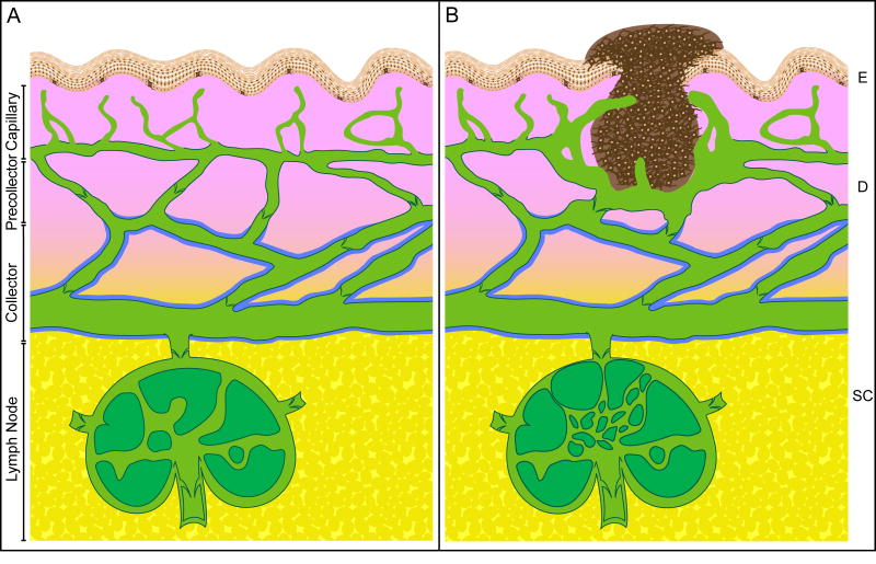
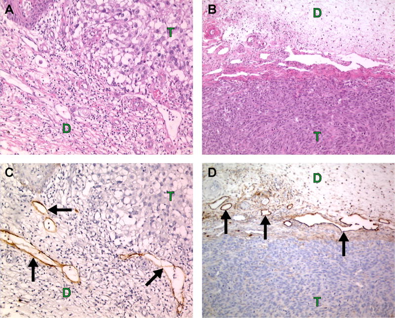
Physiologically, the lymphatic system regulates fluid volume in the interstitium and provides a conduit for immune cells to travel to lymph nodes, but pathologically, the lymphatic system serves as a primary escape route for cancer cells. Lymphatic capillaries have a thin discontinuous basement membrane, lack pericyte coverage and often contain endothelial cell gaps that can be invaded by immune cells (or tumor cells). In addition, tumor cells and stromal cells in the tumor microenvironment secrete factors that stimulate lymphangiogenesis, the growth of lymphatic endothelial cells and the sprouting of lymphatic capillaries. As a result, many tumors are surrounded by large, hyperplastic, peri-tumoral lymphatic vessels and less frequently are invaded by intra-tumoral lymphatic vessels. Carcinoma cells commonly metastasize through these lymphatic vessels to regional lymph nodes. The presence of metastatic cells in the sentinel lymph node is a prognostic indicator for many types of cancer, and the degree of dissemination determines the therapeutic course of action. Lymphangiogenesis is currently at the frontier of metastasis research. Recent strides in this field have uncovered numerous signaling pathways specific for lymphatic endothelial cells and vascular endothelial cells. This review will provide an overview of tumor lymphangiogenesis and current strategies aimed at inhibiting lymphatic metastasis. Novel therapeutic approaches that target the tumor cells as well as the vascular and lymphatic endothelial compartments are discussed.
Figures
Similar articles
-
Suppression of tumor lymphangiogenesis and lymph node metastasis by blocking vascular endothelial growth factor receptor 3 signaling.J Natl Cancer Inst. 2002 Jun 5;94(11):819-25. doi: 10.1093/jnci/94.11.819. J Natl Cancer Inst. 2002. PMID: 12048269
-
Molecular control of lymphatic metastasis.Ann N Y Acad Sci. 2008;1131:225-34. doi: 10.1196/annals.1413.020. Ann N Y Acad Sci. 2008. PMID: 18519975 Review.
-
Tumor and lymph node lymphangiogenesis--impact on cancer metastasis.J Leukoc Biol. 2006 Oct;80(4):691-6. doi: 10.1189/jlb.1105653. Epub 2006 Jun 22. J Leukoc Biol. 2006. PMID: 16793912 Review.
-
Lymphatic vessel activation in cancer.Ann N Y Acad Sci. 2008;1131:235-41. doi: 10.1196/annals.1413.021. Ann N Y Acad Sci. 2008. PMID: 18519976 Review.
-
Tumor necrosis factor superfamily 15 promotes lymphatic metastasis via upregulation of vascular endothelial growth factor-C in a mouse model of lung cancer.Cancer Sci. 2018 Aug;109(8):2469-2478. doi: 10.1111/cas.13665. Epub 2018 Jul 20. Cancer Sci. 2018. PMID: 29890027 Free PMC article.
Cited by
-
Receptor tyrosine kinases: biological functions and anticancer targeted therapy.MedComm (2020). 2023 Dec 7;4(6):e446. doi: 10.1002/mco2.446. eCollection 2023 Dec. MedComm (2020). 2023. PMID: 38077251 Free PMC article. Review.
-
Regulation of epithelial plasticity by miR-424 and miR-200 in a new prostate cancer metastasis model.Sci Rep. 2013 Nov 6;3:3151. doi: 10.1038/srep03151. Sci Rep. 2013. PMID: 24193225 Free PMC article.
-
VEGF-D-induced draining lymphatic enlargement and tumor lymphangiogenesis promote lymph node metastasis in a xenograft model of ovarian carcinoma.Reprod Biol Endocrinol. 2014 Feb 6;12:14. doi: 10.1186/1477-7827-12-14. Reprod Biol Endocrinol. 2014. PMID: 24502459 Free PMC article.
-
Lymphangiogenesis and metastasis--a closer look at the neuropilin/semaphorin3 axis.Microvasc Res. 2014 Nov;96:68-76. doi: 10.1016/j.mvr.2014.07.006. Epub 2014 Aug 1. Microvasc Res. 2014. PMID: 25087623 Free PMC article. Review.
-
The Contribution of Angiogenesis to the Process of Metastasis.Cancer J. 2015 Jul-Aug;21(4):267-73. doi: 10.1097/PPO.0000000000000138. Cancer J. 2015. PMID: 26222078 Free PMC article. Review.
References
-
- Abuharbeid S, Czubayko F, Aigner A. The fibroblast growth factor-binding protein FGF-BP. Int J Biochem Cell Biol. 2006;38:1463–8. - PubMed
-
- Achen MG, Williams RA, Baldwin ME, Lai P, Roufail S, Alitalo K, Stacker SA. The angiogenic and lymphangiogenic factor vascular endothelial growth factor-D exhibits a paracrine mode of action in cancer. Growth Factors. 2002;20:99–107. - PubMed
-
- Achen MG, Roufail S, Domagala T, Catimel B, Nice EC, Geleick DM, Murphy R, Scott AM, Caesar C, Makinen T, Alitalo K, Stacker SA. Monoclonal antibodies to vascular endothelial growth factor-D block its interactions with both VEGF receptor-2 and VEGF receptor-3. Eur J Biochem. 2000;267:2505–15. - PubMed
-
- Ahmed SI, Thomas AL, Steward WP. Vascular endothelial growth factor (VEGF) inhibition by small molecules. J Chemother. 2004;16 4:59–63. - PubMed
Publication types
MeSH terms
Substances
Grants and funding
LinkOut - more resources
Full Text Sources
Research Materials
