. 2008 Jan;9(1):11-9.
doi: 10.1016/j.jpain.2007.07.008. Epub 2007 Oct 24.

Decreased substance P and NK1 receptor immunoreactivity and function in the spinal cord dorsal horn of morphine-treated neonatal rats

Affiliations

Decreased substance P and NK1 receptor immunoreactivity and function in the spinal cord dorsal horn of morphine-treated neonatal rats

Lisa M Thomson et al. J Pain. 2008 Jan.

Abstract

Opiate analgesic tolerance is defined as a need for higher doses of opiates to maintain pain relief after prolonged opiate exposure. Though changes in the opioid receptor undoubtedly occur during conditions of opiate tolerance, there is increasing evidence that opiate analgesic tolerance is also caused by pronociceptive adaptations in the spinal cord. We have previously observed increased glutamate release in the spinal cord dorsal horn of neonatal rats made tolerant to the opiate morphine. In this study, we investigate whether spinal substance P (SP) and its receptor, the neurokinin 1 (NK1) receptor, are also modulated by prolonged morphine exposure. Immunocytochemical studies show decreased SP- and NK1-immunoreactivity in the dorsal horn of morphine-treated rats, whereas SP mRNA in the dorsal root ganglia is not changed. Electrophysiological studies show that SP fails to activate the NK1 receptor in the morphine-treated rat. Taken together, the data indicate that chronic morphine treatment in the neonatal rat is characterized by a loss of SP effects on the NK1 receptor in lamina I of the neonatal spinal cord dorsal horn. The results are discussed in terms of compensatory spinal cord processes that may contribute to opiate analgesic tolerance.

Perspective: This article describes anatomical and physiological changes that occur in the spinal cord dorsal horn of neonatal rats after chronic morphine treatment. These changes may represent an additional compensatory process of morphine tolerance and may represent an additional therapeutic target for the retention and restoration of pain relief with prolonged morphine treatment.


Figures
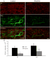
Figure 1
Figure 1
SP- and NK1-ir are significantly reduced in morphine-treated rats. A. Confocal micrographs of SP- and NK1-ir in lumbar dorsal horn of morphine-treated and control rats. SP- (purple) and NK1- (green) immunoreactivity were reduced in the outer laminae of the spinal cord in morphine-treated rats. The overlapping distribution of both markers is shown in the bottom panels. Scale bars = 10 μm. B. The average weighted intensity of SP- (left) and NK1-ir (right) in control (black bars) and morphine-treated (gray bars) rats (mean ± SEM). *,*** represent significant differences from control rats (P<0.05, P <0.01, respectively; Mann-Whitney rank sum test; averaged weighted intensities were calculated for 38 cells from morphine–treated rats and 34 cells from control rats; n= 3 rats).
Figure 2
Figure 2
SP induces a reversible potentiation of sEPSC frequency in lamina I spinal cord dorsal horn cells from control, but not morphine-treated rats. A. Representative recordings from a lamina I cell from a control (top) and a morphine-treated (bottom) rat following the application of SP (1 μM). B. Average effect of SP on the rate of spontaneous excitatory postsynaptic currents (sEPSCs) in the spinal cord dorsal horn of control and morphine-treated rats (mean ± SEM). SP (black bars) induces a reversible potentiation of sEPSC rate in lamina I cells from control (n=10 cells for SP and Wash), but not morphine-treated (n=11 cells for SP and Wash) rats. * indicates significant difference from washout of SP (Wash, gray bars, P < 0.05, Repeated Measures ANOVA with Fisher’s LSD post-hoc comparisons). C. Application of L732,138 (L732, 4 μM, filled bars), a selective antagonist of the NK1 receptor, does not affect sEPSC rate in lamina I spinal cord dorsal horn cells of either control (n= 5, 4 cells for L732, Wash (gray bars) respectively) or morphine-treated rats (n = 7 cells for L732 and Wash) (P> 0.05, Repeated Measures ANOVA).
Figure 3
Figure 3
SP depolarizes lamina I cells in the spinal cord dorsal horn of control, but not morphine-treated rats. A. Representative trace of holding current of a lamina I cell from a control (top) and morphine-treated (bottom) rat. SP (1 μM) causes an inward shift in holding current in a lamina I cell from a control rat (approximately 1 ½ to 2 min from SP application), but causes no change in holding current from a lamina I cell from a morphine-treated rat. B. The average changes in holding current (pA; mean ± SEM) from baseline show that SP (black bars) induces a reversible increase in negative holding current in lamina I cells from control rats (n= 9, 8 cells for SP and Wash, respectively) but not lamina I cells from morphine-treated rats (n=10, 9 cells for SP and Wash, respectively). ˆˆ indicates significant difference from morphine-treated cells following SP application (P <0.01, Repeated Measures ANOVA with Fisher’s LSD post-hoc comparisons); ** indicates significant difference from washout of SP (Wash, gray bars, P < 0.01, Repeated Measures ANOVA with Fisher’s LSD post-hoc comparisons).
Figure 4
Figure 4
SP does not activate NK1 receptors on lamina I cells of morphine-treated rats across a range of voltage steps. A. Protocol used to evaluate the effect of SP (1μM) on current across a range of voltage steps (-140 to 20 mV). B. Representative IV curve before (baseline, filled diamonds) and 2 min after SP application (open squares) in a lamina I cell from a control rat. SP augments inward currents at hyperpolarized potentials (-140 to −80 mV) and outward currents at depolarized potentials (-60 to 20 mV). C. Representative IV curve before (baseline, filled diamonds) and 2 min after SP application (open squares) in a lamina I cell from a morphine-treated rat. SP causes no change in current at any tested voltage step. D. Grouped data showing the effect of SP on current in lamina I cells from control and morphine-treated rats. The SP effect was measured by subtracting the current after SP application from the baseline current before SP application at each voltage step for all recorded cells. *, ** indicate significant difference from lamina I cells from morphine-treated rats (P < 0.05 and P<0.01 respectively, Repeated Measures ANOVA with Fisher’s LSD post-hoc comparisons, n = 4 control cells (filled diamonds), 6 morphine-treated cells (open squares)).

References

    1. Abbadie C, Brown JL, Mantyh PW, Basbaum AI. Spinal cord substance P receptor immunoreactivity increases in both inflammatory and nerve injury models of persistent pain. Neuroscience. 1996;70:201–9. - PubMed
    1. Afrah AW, Fiska A, Gjerstad J, Gustafsson H, Tjolsen A, Olgart L, Stiller CO, Hole K, Brodin E. Spinal substance P release in vivo during the induction of long-term potentiation in dorsal horn neurons. Pain. 2002;96:49–55. - PubMed
    1. Aosaki T, Kawaguchi Y. Actions of substance P on rat neostriatal neurons in vitro. J Neurosci. 1996;16:5141–53. - PMC - PubMed
    1. Belanger S, Ma W, Chabot JG, Quirion R. Expression of calcitonin gene-related peptide, substance P and protein kinase C in cultured dorsal root ganglion neurons following chronic exposure to mu, delta and kappa opiates. Neuroscience. 2002;115:441–53. - PubMed
    1. Berger AJ, Takahashi T. Serotonin enhances a low-voltage-activated calcium current in rat spinal motoneurons. J Neurosci. 1990;10:1922–8. - PMC - PubMed

Publication types

MeSH terms

LinkOut - more resources