. 2007 Dec;189(24):9101-7.
doi: 10.1128/JB.01336-07. Epub 2007 Oct 19.

Crystal structure of the NADH:quinone oxidoreductase WrbA from Escherichia coli

Affiliations

Crystal structure of the NADH:quinone oxidoreductase WrbA from Escherichia coli

Susana L A Andrade et al. J Bacteriol. 2007 Dec.

Abstract

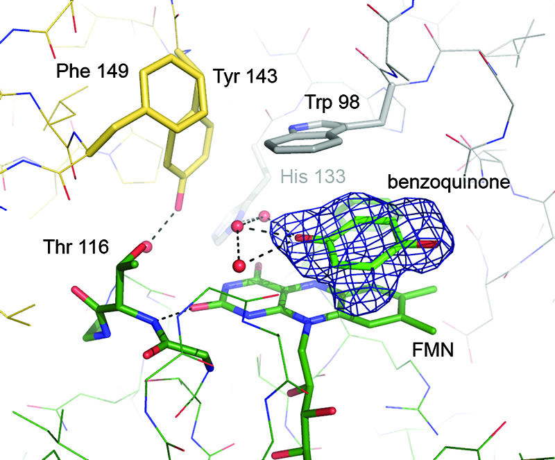
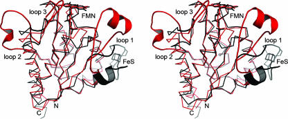
The flavoprotein WrbA, originally described as a tryptophan (W) repressor-binding protein in Escherichia coli, has recently been shown to exhibit the enzymatic activity of a NADH:quinone oxidoreductase. This finding points toward a possible role in stress response and in the maintenance of a supply of reduced quinone. We have determined the three-dimensional structure of the WrbA holoprotein from E. coli at high resolution (1.66 A), and we observed a characteristic, tetrameric quaternary structure highly similar to the one found in the WrbA homologs of Deinococcus radiodurans and Pseudomonas aeruginosa. A similar tetramer was originally observed in an iron-sulfur flavoprotein involved in the reduction of reactive oxygen species. Together with other, recently characterized proteins such as YhdA or YLR011wp (Lot6p), these tetrameric flavoproteins may constitute a large family with diverse functions in redox catalysis. WrbA binds substrates at an active site that provides an ideal stacking environment for aromatic moieties, while providing a pocket that is structured to stabilize the ADP part of an NADH molecule in its immediate vicinity. Structures of WrbA in complex with benzoquinone and NADH suggest a sequential binding mechanism for both molecules in the catalytic cycle.


Figures

FIG. 1.
FIG. 1.
Crystals of E. coli WrbA. The tetragonal bipyramidal shape of the crystals reflects their P422 symmetry.
FIG. 2.
FIG. 2.
Cartoon representation of WrbA from E. coli. The stereo image of the protein chain shows the N terminus in blue and the C terminus in red. Strands of the central, parallel β-sheet and the surrounding α-helices are numbered according to their occurrence in the protein sequence. The FMN cofactor is bound peripherally at the C-terminal end of the β-sheet.
FIG. 3.
FIG. 3.
Close-up of the active site of E. coli WrbA. Residues from three monomers combine to create a specific, hydrophobic active-site pocket. Aromatic substrates can be stacked in between the FMN moiety and the side chain of Trp 98. All residues are colored by chain.
FIG. 4.
FIG. 4.
Tetrameric arrangement of WrbA and binding of NADH. (A) Cartoon representation of the tetramer. A single monomer is shown, colored as described in the legend of Fig. 1. Note the extended loop 2 region (Fig. 2) interacting with a neighboring monomer. (B) Surface representation of the tetramer, centered on the active site of one monomer. The picture shows a PEG molecule bound at the active site, an NADH molecule in close proximity, and several further PEG molecules along a dimer interface. (C) Stereo representation of a close-up of the active site in the same orientation as in panel B. The left and right images are for the left and right eyes, respectively. The nicotinamide ring of NADH stacks onto the side chain of Tyr 12.
FIG. 5.
FIG. 5.
Binding of benzoquinone to the FMN site of WrbA. The picture shows the amino acid residues surrounding the active site and an FoFc density map, contoured at 3.5 σ, from which the benzoquinone molecule was omitted.
FIG. 6.
FIG. 6.
Superposition of the monomers of E. coli WrbA (red) and M. thermophila ISF (black). Based on a common flavodoxin-like fold, the structures differ in three characteristic loop regions. Strongest variations are visible in the loop region 1, where ISF binds a [4Fe:4S] cluster while WrbA forms a specific binding cleft for NADH at a very different position in the monomer. However, multimer formation then places both putative electron donor sites at very similar positions in respect to the FMN cofactor.

References

    1. Andrade, S. L. A., F. Cruz, C. L. Drennan, V. Ramakrishnan, D. C. Rees, J. G. Ferry, and O. Einsle. 2005. Structures of the iron-sulfur flavoproteins from Methanosarcina thermophila and Archaeoglobus fulgidus. J. Bacteriol. 187:3848-3854. - PMC - PubMed
    1. Brock, B. J., and M. H. Gold. 1996. 1,4-benzoquinone reductase from the basidiomycete Phanerochaete chrysosporium: spectral and kinetic analysis. Arch. Biochem. Biophys. 331:31-40. - PubMed
    1. Brock, B. J., S. Rieble, and M. H. Gold. 1995. Purification and characterization of a 1,4-benzoquinone reductase from the basidiomycete Phanerochaete chrysosporium. Appl. Environ. Microbiol. 61:3076-3081. - PMC - PubMed
    1. Brünger, A. T. 1993. Assessment of phase accuracy by cross validation: the free R-value. Methods and applications. Acta Crystallogr. D 49:24-36. - PubMed
    1. Cohen, R., M. R. Suzuki, and K. E. Hammel. 2004. Differential stress-induced regulation of two quinone reductases in the brown rot basidiomycete Gloeophyllum trabeum. Appl. Environ. Microbiol. 70:324-331. - PMC - PubMed

Publication types

MeSH terms

LinkOut - more resources