. 2007 Dec;6(12):2365-75.
doi: 10.1128/EC.00275-07. Epub 2007 Oct 19.

Establishment of the ambient pH signaling complex in Aspergillus nidulans: PalI assists plasma membrane localization of PalH

Affiliations

Establishment of the ambient pH signaling complex in Aspergillus nidulans: PalI assists plasma membrane localization of PalH

Ana M Calcagno-Pizarelli et al. Eukaryot Cell. 2007 Dec.

Abstract

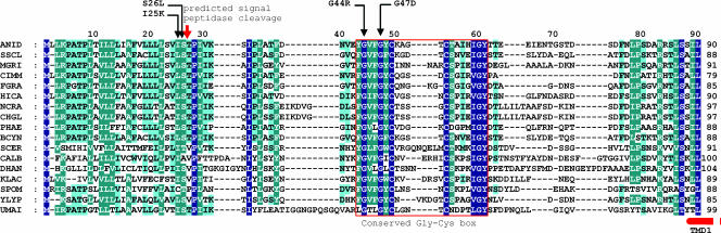
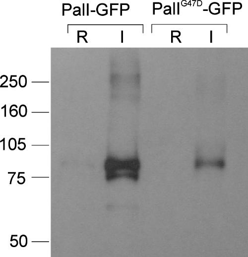
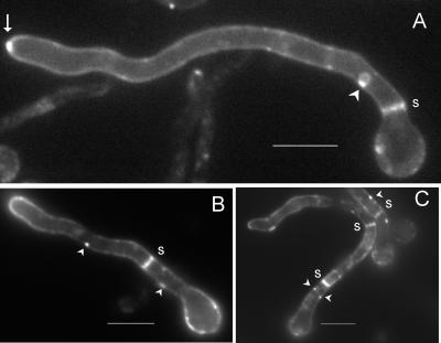
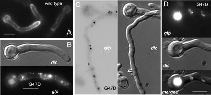
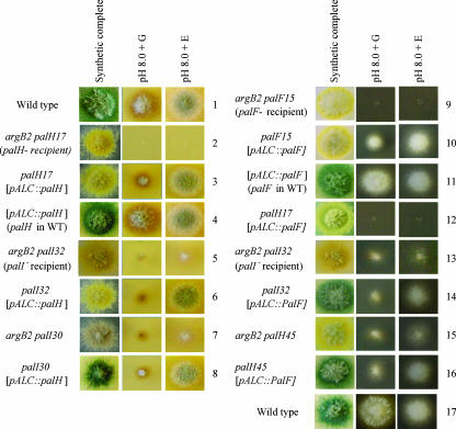
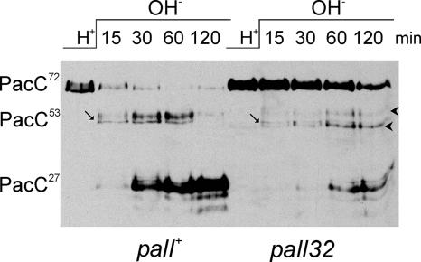
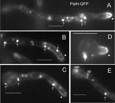
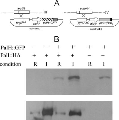
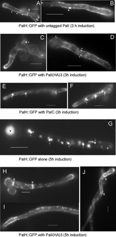
The Aspergillus nidulans ambient pH signaling pathway involves two transmembrane domain (TMD)-containing proteins, PalH and PalI. We provide in silico and mutational evidence suggesting that PalI is a three TMD (3-TMD) protein with an N-terminal signal peptide, and we show that PalI localizes to the plasma membrane. PalI is not essential for the proteolytic conversion of the PacC translation product into the processed 27-kDa form, but its absence markedly reduces the accumulation of the 53-kDa intermediate after cells are shifted to an alkaline pH. PalI and its homologues contain a predicted luminal, conserved Gly-Cys-containing motif that distantly resembles a Gly-rich dimerization domain. The Gly44Arg and Gly47Asp substitutions within this motif lead to loss of function. The Gly47Asp substitution prevents plasma membrane localization of PalI-green fluorescent protein (GFP) and leads to its missorting into the multivesicular body pathway. Overexpression of the likely ambient alkaline pH receptor, the 7-TMD protein PalH, partially suppresses the null palI32 mutation. Although some PalH-GFP localizes to the plasma membrane, it predominates in internal membranes. However, the coexpression of PalI to stoichiometrically similar levels results in the strong predominance of PalH-GFP in the plasma membrane. Thus, one role for PalI, but possibly not the only role, is to assist with plasma membrane localization of PalH. These data, considered along with previous reports for both Saccharomyces cerevisiae and A. nidulans, strongly support the prevailing model of pH signaling involving two spatially segregated complexes: a plasma membrane complex containing PalH, PalI, and the arrestin-like protein PalF and an endosomal membrane complex containing PalA and PalB, to which PacC is recruited for its proteolytic activation.


Figures

FIG. 1.
FIG. 1.
The N-terminal region of PalI and Rim9p family members. Multiple sequence alignment of the ∼100 N-terminal residues of PalI/Rim9p homologues in filamentous and yeast ascomycetes and the dimorphic basidiomycete Ustilago maydis. Single-residue substitutions of residues within this region that led to complete or partial loss of function are indicated (Table 2). The position of the signal peptide cleavage site predicted by SignalPeptide 3.0 (http://www.cbs.dtu.dk/services/SignalP/) with a 0.84 probability is indicated by a red arrow. The red box area is the conserved Gly-Cys motif. The start of the first TMD, corresponding to the second hydrophobic region from the N termini of the preproteins (10), is indicated by a broken bar. Conserved residues (according to the Blosum62 matrix) were shaded with navy blue, blue-green, and blue, indicating full, more than 80% but less than full conservation, and more than 60% but less than 80% conservation, respectively. ANID, A. nidulans; SSCL, Sclerotinia sclerotiorum; MGRI, Magnaporthe grisea; CIMM, Coccidioides immitis; FGRA, Fusarium graminearum; HICA, Histoplasma capsulatum; NCRA, Neurospora crassa; CHGL, Chaetomium globosum; PHAE, Phaeosphaeria nodorum; BCYN, Botrytis cinerea; SCER, S. cerevisiae RIM9p; CALB, Candida albicans Rim9; DHAN, Debaryomyces hansenii; KLAC, Kluyveromyces lactis; SPOM, Schizosaccharomyces pombe; YLYP, Yarrowia lipolytica; UMAI, Ustilago maydis.
FIG. 2.
FIG. 2.
Western blotting analysis of cells expressing PalI-GFP and PalIG47D-GFP fusion proteins. Strains expressing wild-type and Gly47Asp PalI-GFP fusion proteins under the control of the alcAp promoter were cultured overnight in minimal medium with 0.05% (wt/vol) glucose and shifted for 3 h to either 1% ethanol-containing medium (I, inducing conditions for alcAp) or 1% glucose-containing medium (R, repressing conditions for alcAp) before proceeding to membrane protein extraction. Proteins were analyzed by Western blotting, which was reacted using a cocktail of monoclonal anti-GFP antibodies (Roche). Approximately equal loading of the different lanes was confirmed after protein staining of a duplicate gel. Standards (at left) are in kDa.
FIG. 3.
FIG. 3.
PalI localizes to the plasma membrane. Epifluorescence microscopy using a filter set specific for GFP of germlings cultured in acidic WMM. (A) With ethanol as the sole carbon source. The arrow indicates strong labeling in the position corresponding to the Spitzenkörper. (B and C) Germlings germinated in 0.02% glucose and shifted to WMM with 1% ethanol for 3 h. Membrane-associated punctate structures are indicated by arrowheads, whereas septae are indicated by s. Bars, 5 μm.
FIG. 4.
FIG. 4.
PalIG47D-GFP mislocalization to the endosomal system and the vacuole. (A) GFP fluorescence microscopy of a strain expressing wild-type PalI-GFP. (B, C, and D) Fluorescence microscopy of germlings expressing PalIG47D-GFP, including Nomarski differential interference contrast (dic) and GFP images, as indicated. (C) Fluorescent cytosolic dots are shown, using reversed contrast for clarity. (D) Vacuole labeled with GFP fluorescence, as seen in mutant but not in wild-type germlings. Germlings were cultured overnight at 25°C in acidic WMM with 0.02% glucose and shifted for 3 h to the same medium with ethanol as the sole carbon source. Bars, 5 μm.
FIG. 5.
FIG. 5.
Overexpression of PalH and, to a lesser extent, PalF suppresses the reduced ability of palI loss-of-function mutant strains to grow at an alkaline pH. Forced expression of PalH and PalF was driven by single-copy transgenes integrated at the argB locus, using the alcAp promoter. Thus, recipient strains carry the argB2 allele in addition to the relevant pH regulatory mutations (as shown also in Fig. 7), and transgene expression mirrors that of the alcA gene, which is induced by ethanol (E) and repressed by glucose (G). Under conditions of arginine supplementation, the argB2 mutation does not affect growth on pH media. The synthetic complete (pH 6.5) medium is permissive for the acidity-mimicking pal mutants. However, in contrast to the wild type (strains 1, 4, and 11), the pal mutants grow poorly at pH 8.0, unless this phenotype is complemented (control strains 3 and 10) or suppressed (see relevant strains) by expression of the transgene. Growth on alkaline pH plates is the most sensitive test for ambient pH signaling, as shown by the residual growth under repressing conditions of strains 3 and 10, due to very low levels of expression of alcAp under repressing conditions. palH17 and palF15 carry phenotypically null mutations. palH45 is a leaky palH mutation allowing some growth at alkaline pH. As noted in the text, palI mutations characteristically allow some growth at alkaline pH (strains 5, 7, and 13). Note that strains carry different spore color markers (wild-type green, mutant yellow, or mutant chartreuse).
FIG. 6.
FIG. 6.
The complete loss-of-function mutation palI32 does not fully prevent the formation of PacC27 in pH shift experiments. Shown is Western blotting analysis of Myc-tagged PacC in extracts from wild-type or palI32 cells cultured under acidic conditions (H+) and transferred to alkaline conditions (OH) for the indicated time points before proceeding with protein extraction. Arrows indicate (nonphosphorylated) PacC53 (see text), whereas arrowheads indicate the two abnormal bands seen in the mutant but not in the wild type.
FIG. 7.
FIG. 7.
PalH-GFP expressed from the alcAp localizes to the plasma membrane but predominates in cytosolic specks. (A) A germling where PalH-GFP localization at the apical plasma membrane of the shorter germ tube is clearly visible (arrowhead), although PalH-GFP predominates in cytosolic specks (arrows). (B and C) Plasma membrane localization of PalH-GFP (arrowheads) in longer germlings is less prominent than that seen in internal specks (arrows). (D) Very young germling showing clear polarization of PalH-GFP at the apical plasma membrane. (E) PalH-GFP also labels septae (sp) and eventually reaches the lumen of the vacuole (vac). Bars, 5 μm.
FIG. 8.
FIG. 8.
A system for the simultaneous expression of two proteins from single-copy transgenes targeted to argB and pyroA. (A) Targeted integration of the transforming plasmids to the argB and pyroA genes, located at chromosomes III and IV, respectively. Use of the argBBgl2 allele for site-directed integration has been reported previously (31, 39). (B) Western blotting analysis of PalH-GFP and PalI-(HA)3 expression driven by alcAp. Strains carrying (+) or lacking (−) the transgenes are indicated. The PalH-GFP transgene was integrated at argB, whereas that driving expression of PalI-(HA)3 was integrated at pyroA. The top and bottom panels were revealed with anti-GFP and anti-HA antibodies, respectively.
FIG. 9.
FIG. 9.
Coexpression of PalI from the alcAp promoter results in the plasma membrane localization of PalH-GFP. Germlings of strains expressing the indicated proteins under the control of the alcAp gene were cultured as described in the legend to Fig. 3, with a 3-h (A to F) or a 5-h (G to J) induction period, as indicated. (A and B) Coexpression of PalI with PalH-GFP promotes the plasma membrane localization of the latter. (C and D) As described in the legend to panels A and B above, using PalI-(HA)3 rather than untagged PalI. (E and F) Coexpression of PalC does not promote plasma membrane localization of PalH-GFP. Note that the distribution of PalH-GFP in this strain cannot be distinguished from that shown in Fig. 7 (in the absence of PalC coexpression). (G) After a relatively long period of PalH-GFP transgene induction, the reporter almost exclusively localizes to strongly fluorescent cytosolic specks (arrows) and to the vacuole (v). (H, I, and J) In marked contrast, PalH-GFP predominates at the plasma membrane if PalI-(HA)3 is coexpressed using the same induction regimen. Note the clearly patchy appearance of PalH-GFP at the plasma membrane, the strong labeling of septae (s), and the peripheral punctate structures. Bars, 5 μm.

References

    1. Arst, H. N., Jr., E. Bignell, and J. Tilburn. 1994. Two new genes involved in signaling ambient pH in Aspergillus nidulans. Mol. Gen. Genet. 245:787-790. - PubMed
    1. Arst, H. N., Jr., and M. A. Peñalva. 2003. pH regulation in Aspergillus and parallels with higher eukaryotic regulatory systems. Trends Genet. 19:224-231. - PubMed
    1. Babst, M., D. J. Katzmann, E. J. Estepa-Sabal, T. Meerloo, and S. D. Emr. 2002. ESCRT-III: an endosome-associated heterooligomeric protein complex required for MVB sorting. Dev. Cell 3:271-282. - PubMed
    1. Bartnicki-García, S., F. Hergert, and G. Gierz. 1989. Computer simulation of fungal morphogenesis and the mathematical basis for hyphal tip growth. Protoplasma 153:46-57.
    1. Barwell, K. J., J. H. Boysen, W. Xu, and A. P. Mitchell. 2005. Relationship of DFG16 to the Rim101p pH response pathway in Saccharomyces cerevisiae and Candida albicans. Eukaryot. Cell 4:890-899. - PMC - PubMed

Publication types

Substances

LinkOut - more resources