. 2008 Mar 20;27(13):1951-60.
doi: 10.1038/sj.onc.1210832. Epub 2007 Oct 22.

Genome-wide DNA copy number analysis in pancreatic cancer using high-density single nucleotide polymorphism arrays

Affiliations

Genome-wide DNA copy number analysis in pancreatic cancer using high-density single nucleotide polymorphism arrays

T Harada et al. Oncogene. .

Abstract

To identify genomic abnormalities characteristic of pancreatic ductal adenocarcinoma (PDAC) in vivo, a panel of 27 microdissected PDAC specimens were analysed using high-density microarrays representing approximately 116 000 single nucleotide polymorphism (SNP) loci. We detected frequent gains of 1q, 2, 3, 5, 7p, 8q, 11, 14q and 17q (> or =78% of cases), and losses of 1p, 3p, 6, 9p, 13q, 14q, 17p and 18q (> or =44%). Although the results were comparable with those from array CGH, regions of those genetic changes were defined more accurately by SNP arrays. Integrating the Ensembl public data, we have generated 'gene' copy number indices that facilitate the search for novel candidates involved in pancreatic carcinogenesis. Copy numbers in a subset of the genes were validated using quantitative real-time PCR. The SKAP2/SCAP2 gene (7p15.2), which belongs to the src family kinases, was most frequently (63%) amplified in our sample set and its recurrent overexpression (67%) was confirmed by reverse transcription-PCR. Furthermore, fluorescence in situ hybridization and in situ RNA hybridization analyses for this gene have demonstrated a significant correlation between DNA copy number and mRNA expression level in an independent sample set (P<0.001). These findings indicate that the dysregulation of SKAP2/SCAP2, which is mostly caused by its increased gene copy number, is likely to be associated with the development of PDAC.


Figures

Figure 1
Figure 1
The overview of genomic changes of all chromosomes in a total of 27 microdissected PDAC tissues, determined by the Affymetrix 100 K SNP arrays. (see Supplementary Figure 1 for all the details) ‘Genetic gains’ are shown as green bars and ‘losses’ as red bars according to the genomic position (Build 35). Thick bars are used to depict ‘high-level amplifications’ and ‘homozygous deletions’. Blue bars indicate ‘LOH regions’ and grey, thick bars are used for ‘UPD regions’.
Figure 2
Figure 2
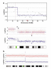
(a) Comparison of the results between CGH and SNP arrays. Upper: Chromosome 18 in PC16, determined by array CGH (see ref. Harada et al., 2007). The blue line is used to depict the smoothed DCN values. Lower: The same sample was analysed by SNP arrays, showing more distinct physical boundaries that enabled to identify small size of DCN alterations (asterisks). (b) ‘High-level amplification’ detected by SNP arrays (chromosome 11 in PC33). This amplicon size was approximately 1.35 Mb.
Figure 3
Figure 3
Comparison of DCN alterations identified by SNP arrays (blue columns) and q-PCR (violet columns) analyses. A significant correlation (r=0.72) was observed between two data sets. All the data are shown in Supplementary Table 3.
Figure 4
Figure 4
(A) The minimal common region (7p15.2) of 7p gain, defined by DCN analysis of 27 PDAC cases. The green bars below the chromosome define the region of copy number gain in each case analysed in this study. The approximately 1 Mb region, which was most frequently gained in our sample set, was surrounded by black dots. The genes contained in the minimal region at 7p15.2 are listed below. SNP markers spotted on Affymetrix 100 K arrays are shown at the bottom. (B) Transcript levels of five candidate genes within the minimal region in normal pancreas and PDAC tissues, determined by RT–PCR. Samples were run in the following order: lane 1–2, normal pancreas; lane 3–14, PDACs; lane 15, negative control. Among five genes, the only SCAP2 transcript was not detectable in normal pancreas tissues, whereas it was upregulated in eight out of 12 PDAC tissues (67%). (C) The FISH results in two representative cases. (a) No copy number change of SCAP2 in PC58 and (b) a genetic gain in PC63. Original magnification: × 1000. (D) SCAP2 mRNA expression in normal epithelial cells and PDAC cells, determined by ISH. (a) No signals (score 0) were detected in ductal (arrow) and islet cells (arrow heads) of normal pancreas, whereas very subtle, patchy signals were observed in acinar cells. (b) ISH conducted with a sense SCAP2 riboprobe, used as a negative control. Arrow indicates ductal cells of normal pancreas. (c) Strong signal (score 2) in moderately differentiated PDAC cells (PC53). (d) Higher level of expression (score 1) in metastatic PDAC cells (PC66) compared to normal hepatic cells (asterisk). Arrows indicate two micrometastatic lesions. Original magnification: (a and b), × 400; (c), × 100; (d), × 40.

References

    1. Andersen CL, Wiuf C, Kruhoffer M, Korsgaard M, Laurberg S, Orntoft TF. Frequent occurrence of uniparental disomy in colorectal cancer. Carcinogenesis. 2007;28:38–48. - PubMed
    1. Bardeesy N, DePinho RA. Pancreatic cancer biology and genetics. Nat Rev Cancer. 2002;2:897–909. - PubMed
    1. Bignell GR, Huang J, Greshock J, Watt S, Butler A, West S, et al. High-resolution analysis of DNA copy number using oligonucleotide microarrays. Genome Res. 2004;14:287–295. - PMC - PubMed
    1. Buchholz M, Braun M, Heidenblut A, Kestler HA, Kloppel G, Schmiegel W, et al. Transcriptome analysis of microdissected pancreatic intraepithelial neoplastic lesions. Oncogene. 2005;24:6626–6636. - PubMed
    1. Calhoun ES, Hucl T, Gallmeier E, West KM, Arking DE, Maitra A, et al. Identifying allelic loss and homozygous deletions in pancreatic cancer without matched normals using high-density single-nucleotide polymorphism arrays. Cancer Res. 2006;66:7920–7928. - PubMed

Publication types

MeSH terms

Substances