Review
. 2007;49(3):165-81.
doi: 10.1007/s12013-007-9001-4. Epub 2007 Oct 2.

A comparison of the mechanical and structural properties of fibrin fibers with other protein fibers

Affiliations
Review

A comparison of the mechanical and structural properties of fibrin fibers with other protein fibers

M Guthold et al. Cell Biochem Biophys. 2007.

Abstract

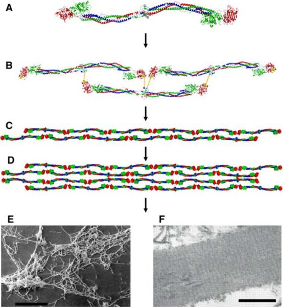
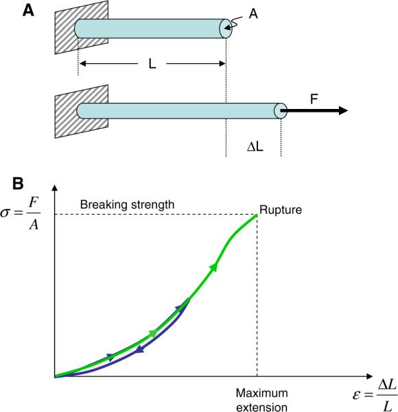
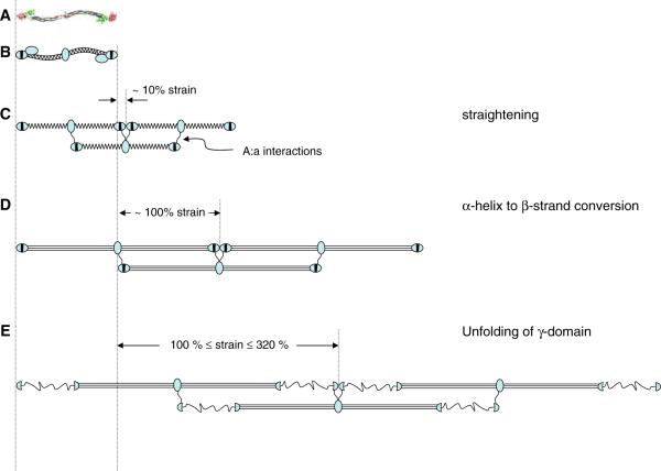
In the past few years a great deal of progress has been made in studying the mechanical and structural properties of biological protein fibers. Here, we compare and review the stiffness (Young's modulus, E) and breaking strain (also called rupture strain or extensibility, epsilon(max)) of numerous biological protein fibers in light of the recently reported mechanical properties of fibrin fibers. Emphasis is also placed on the structural features and molecular mechanisms that endow biological protein fibers with their respective mechanical properties. Generally, stiff biological protein fibers have a Young's modulus on the order of a few Gigapascal and are not very extensible (epsilon(max) < 20%). They also display a very regular arrangement of their monomeric units. Soft biological protein fibers have a Young's modulus on the order of a few Megapascal and are very extensible (epsilon(max) > 100%). These soft, extensible fibers employ a variety of molecular mechanisms, such as extending amorphous regions or unfolding protein domains, to accommodate large strains. We conclude our review by proposing a novel model of how fibrin fibers might achieve their extremely large extensibility, despite the regular arrangement of the monomeric fibrin units within a fiber. We propose that fibrin fibers accommodate large strains by two major mechanisms: (1) an alpha-helix to beta-strand conversion of the coiled coils; (2) a partial unfolding of the globular C-terminal domain of the gamma-chain.


Figures

Fig. 1
Fig. 1
Crystal structure of chicken fibrinogen (7) and fiber assembly. (A) The fibrinogen molecule is 46 nm long and 4.5 nm in diameter and consists of six polypeptide chains; two α-chains (blue), two β-chains (green), and two γ-chains (red). (B) The monomers assemble in a half-staggered fashion to form the two-stranded protofibrils (C); the A: a interactions are shown as yellow lines. (D) The protofibrils aggregate laterally to form thicker fibers, branching occurs and eventually a full clot (E) is formed in which fiber diameters range from about 20 to 200 nm ((E) shows an SEM image of a 50% lysed thrombus containing fibrin fibers and platelets; courtesy R. R. Hantgan; reproduced with permission from [8]; scale bar: 10 μm). (F) A striated pattern with a periodicity of 22.5 nm (half the length of a fibrin monomer) is seen in the TEM image of a longitudinal cross section of a single fiber (Image: W. Liu, scale bar 200 nm). The striated pattern is due to the half-staggered arrangement of the monomers
Fig. 2
Fig. 2
Stress–strain curves of stretched fibers. (A) A force F is applied in the longitudinal direction to a fiber with length L and cross sectional area A. The fiber extends by an amount ΔL. (B) A schematic stress–strain curve of the stretching of a fiber. The slope of the curve corresponds to the stiffness of the fiber. In a linear, elastic model, the stiffness (slope) is the called Young's modulus of the material. The maximum extension at which the fiber ruptures is called breaking strain (or extensibility) of the fiber
Fig. 3
Fig. 3
Experimental set-up to determine fibrin fiber breaking strain (extensibility) (Figure adopted from [1] with permission). The experimental set-up to stretch single fibrin fibers is depicted photographically in Fig. 2A and schematically in Fig. 2B. The atomic force microscope (AFM) was used to stretch fibers that were suspended across ~12 μm-wide channels; the fluorescence microscope was used to record movies of this stretching process. This set-up has several advantageous features for stretching out fibers. Somewhat fortuitously, we found that the fibers were very well anchored on the ridges of the striated substrate as the anchoring points rarely changed; even at the most extreme fiber extensions. This implies that the observed deformations are not due to fiber slipping on the ridges. We selected fibers that bridged the grooves at a right angle with respect to the ridge edge. Thus, our set-up yielded a well-defined, easy-to-analyze geometry. By suspending the fibers over grooves, the substrate did not interfere with the measurement. Being able to record movies of the stretching process allowed us to accurately determine the lengths of the fibers and ascertain that the fibers did not slip at the anchoring points. The technique may also be applied to other fibers
Fig. 4
Fig. 4
Extensibility of fibrin fibers (adopted from [1] with permission and modified). (A–F) Snapshots from a fluorescence microscopy movie of a fiber extensibility experiment (crosslinked batroxobin fiber). A fluorescently labeled fiber is suspended between two ridges that appear as two horizontal bands. The AFM cantilever is seen from underneath as a dark, ~35 μm-wide rectangle; the location of the AFM tip is indicated by a white dot. The AFM tip is moved to the right, thus stretching the fiber; the lower segment of the fiber breaks at 183% strain and the upper segment of the fiber breaks at 278%. Histogram (G) and bar graph (H) representations of the extensibilities of the four different types of fibrin fibers; Thr = thrombin, Bat = Batroxobin, X = crosslinking
Fig. 5
Fig. 5
Elastic limit of fibrin fibers (adopted from [1] with permission and modified). (A–D) A fibrin fiber (thrombin, crosslinked) was strained 80% (C), from which it snapped back to its original length (D) without any permanent lengthening. (D–G) The same fiber was strained to 230% (F); at this strain it did suffer some permanent damage as the fiber is significantly deformed in image (G). (H) Plot of the amount of permanent deformations (% length increase) upon the release of force versus strain. Data points along the horizontal axis of 0%-permanent length increase indicate elastic deformations. Crosslinked thrombin fibers could be strained over 180% and the other fiber types about 120% without suffering permanent lengthening. None of the fibers analyzed here were ruptured
Fig. 6
Fig. 6
Schematic molecular mechanism models for fibrin fiber extensions. (A) Crystal structure and (B) schematic model of fibrinogen. (C) Half-staggered assembly of three fibrin monomers; molecules are rotated 90° from view in (B); βC domain is no longer depicted; straightening and slightly stretching the molecule may accommodate 10% strain. (D) An α-helix to β-strand conversion of the coiled coil and a slight straightening and alignment of the molecules could accommodate ~100% strain. (E) Higher strains, up to 320% could be accommodated by a partial unfolding of the γC domains; 230% strain is depicted

References

    1. Liu W, Jawerth LM, Sparks EA, Falvo MR, Hantgan RR, Superfine R, Lord ST, Guthold M. Fibrin fibers have extraordinary extensibility and elasticity. Science. 2006;313:634. - PMC - PubMed
    1. Collet JP, Shuman H, Ledger RE, Lee ST, Weisel JW. The elasticity of an individual fibrin fiber in a clot. Proceedings of the National Academy of Sciences of the United States of America. 2005;102:9133. - PMC - PubMed
    1. Weisel JW. The electron-microscope band pattern of hman fibrin—various stains, lateral order, and carbohydrate localization. Journal of Ultrastructure and Molecular Structure Research. 1986;96:176. - PubMed
    1. Hantgan RR, Fowler SB, Erickson HP, Hermans J. Fibrin assembly: A comparison of electron microscopic and light scattering results. Thrombosis and Haemostasis. 1980;44:119. - PubMed
    1. Spraggon G, Everse SJ, Doolittle RF. Crystal structures of fragment D from human fibrinogen and its cross-linked counterpart from fibrin. Nature. 1997;389:455. - PubMed

Publication types

LinkOut - more resources