. 2008 Jan;28(1):131-9.
doi: 10.1128/MCB.01119-07. Epub 2007 Oct 22.

The alpha catalytic subunit of protein kinase CK2 is required for mouse embryonic development

Affiliations

The alpha catalytic subunit of protein kinase CK2 is required for mouse embryonic development

David Y Lou et al. Mol Cell Biol. 2008 Jan.

Abstract

Protein kinase CK2 (formerly casein kinase II) is a highly conserved and ubiquitous serine/threonine kinase that is composed of two catalytic subunits (CK2alpha and/or CK2alpha') and two CK2beta regulatory subunits. CK2 has many substrates in cells, and key roles in yeast cell physiology have been uncovered by introducing subunit mutations. Gene-targeting experiments have demonstrated that in mice, the CK2beta gene is required for early embryonic development, while the CK2alpha' subunit appears to be essential only for normal spermatogenesis. We have used homologous recombination to disrupt the CK2alpha gene in the mouse germ line. Embryos lacking CK2alpha have a marked reduction in CK2 activity in spite of the presence of the CK2alpha' subunit. CK2alpha(-/-) embryos die in mid-gestation, with abnormalities including open neural tubes and reductions in the branchial arches. Defects in the formation of the heart lead to hydrops fetalis and are likely the cause of embryonic lethality. Thus, CK2alpha appears to play an essential and uncompensated role in mammalian development.


Figures

FIG. 1.
FIG. 1.
CK2α targeting. (A) CK2α-targeting vector (II) and targeted allele (III). To generate a CK2α null allele, an 85-bp region of the gene (I) (numbers indicate exons) that included the critical K68 residue (*) in the catalytic ATP-binding pocket was targeted. A 3.7-kb 5′-flanking arm and 4.5-kb 3′-flanking arm were cloned into vector pPNT using KpnI (K), NotI (N), and XhoI. Screening was carried out by PCR using an external flanking and internal neo PCR primer pair (a and b, respectively). (B) To confirm homologous recombination, Southern blotting was carried out following XmnI (X) restriction digestion. A 5′-flanking probe (c) was used to identify the 11-kb wild-type (WT) and 6.1-kb homologously recombined DNA fragments, confirmed with a 3′ probe (d) (not shown). ES cell colonies 7, 24, 30, and 42 were hemizygous for the targeted CK2α allele, while colonies 9, 11, 12, and 32 were wild type. HSV, herpes simplex virus; KO, knockout.
FIG. 2.
FIG. 2.
CK2α−/− embryos lack CK2α mRNA and protein. (A) To detect CK2α mRNA transcripts, E10.5 embryos were genotyped individually by PCR using DNA prepared from the yolk sacs. Three CK2α−/− (−/−) and three CK2α+/+ (+/+) embryos were pooled, and RT-PCR was performed; − indicates CK2α+/+ RNA without RT. (B) To assay for CK2α protein, E10.5 embryos were genotyped, and protein extracts were prepared. Fifteen micrograms of protein from individual embryos was resolved by 10% sodium dodecyl sulfate-polyacrylamide gel electrophoresis and immunoblotted using an anti-CK2α/CK2α′ antibody. Wild-type CK2α+/+ (+/+) embryos contained a major immunoreactive band at ∼42 kDa, consistent with CK2α, and a weaker band at ∼38 kDa, consistent with CKα′. In the knockout (KO) CK2α−/− (−/−) embryos, the intensity of the CK2α′ band was unchanged, and the CK2α band was absent. Heterozygote CK2α+/− (+/−) embryos reproducibly had a CK2α band of half the intensity of the wild type (WT). Tubulin was used as a loading control.
FIG. 3.
FIG. 3.
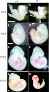
Comparison of wild-type CK2α+/+ (A, C, E, and G) and knockout CK2α−/− (B, D, F, and H) embryos from E8.5 to E11.5; white scale bars (500 μm) indicate the relative sizes of embryos between developmental stages. At E8.5 (A and B), the neural folds in the CK2α−/− embryos failed to elevate like those of CK2α+/+ littermates. By E9.5 (C and D), CK2α−/− hearts were larger than those of their littermates; CK2α−/− neural tubes failed to fuse at the cranial level. At E10.5 (E and F), CK2α−/− embryos were usually smaller, hearts were dilated, and head shape was abnormal. At E11.5 (G and H), CK2α−/− embryos were runted, hearts were no longer were beating, and blood could be seen hemorrhaging into the cranium and thorax.
FIG. 4.
FIG. 4.
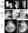
Details of developmental defects in CK2α−/− embryos. (A to C) At E8.5, CK2α−/− neural folds failed to elevate (A). At E9.5, the neural folds failed to fuse along the anterior-posterior axis (arrows indicate the unfused neural tube on the ventral side [B] or dorsal side [C]). (D to F) By E9.5, wild-type embryos have developed the second branchial arch (D) (inset is enlarged in panel E), but it was smaller in most of the CK2α−/− embryos (F) (Table 2). (G and H) At E11, the pericardial sacs of the CK2α−/− embryos were edematous (arrow) due to heart failure, i.e., hydrops fetalis.
FIG. 5.
FIG. 5.
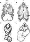
Histological abnormalities in CK2α−/− embryos. Representative E10.5 CK2α+/+ and CK2α−/− embryos were fixed and sectioned through the cranial (A and B) and thoracic (C and D) regions. In the CK2α+/+ head (A), the neural tube has fused, forming a sealed tube that is expanding in preparation for brain development. In the CK2α−/− embryo, because the neural tube (nt) is open anteriorly in the forebrain (fb) region, the posterior tube has collapsed, interfering with the normal development of the hindbrain (hb). The optic vesicles (opv) and the notochord (nc) appear to be similar, but the otic vesicles (otv) in the CK2α−/− embryo are round and thickened compared to those in the CK2α+/+ embryo, which has a characteristic dorsal extension. (C and D) Whereas the CK2α+/+ heart (ht) is forming chambers and developing trabeculation, the heart of the CK2α−/− embryo is still an open tube.
FIG. 6.
FIG. 6.
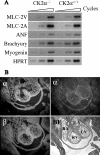
mRNA expression in the embryos. (A) Semiquantitative RT-PCR was used to demonstrate the presence of mRNA encoding markers of mesoderm and cardiac formation in CK2α−/− and CK2α+/+ embryos at E9.5. The triangles indicate increasing numbers of amplification cycles. Transcript levels of the cardiac markers MLC-2V, MLC-2A, and ANF and brachyury and myogenin, markers of mesoderm formation, were measured; hypoxanthine phosphoribosyltransferase (HPRT) was used as an mRNA control. (B) In situ hybridization for CK2 subunit transcripts demonstrates strong relative expression of CK2α mRNA in the ventricles at E13.5, while CK2α′ and CK2β appear to have a more uniform expression throughout the embryo. Sense probes showed no signal and are omitted, and a bright-field (BF) image is shown for orientation, with the right atrium (RA), left atrium (LA), right ventricle (RV), and left ventricle (LV) labeled.

References

    1. Ackermann, K., A. Waxmann, C. V. Glover, and W. Pyerin. 2001. Genes targeted by protein kinase CK2: a genome-wide expression array analysis in yeast. Mol. Cell. Biochem. 22759-66. - PubMed
    1. Bidwai, A. P., J. C. Reed, and C. V. Glover. 1995. Cloning and disruption of CKB1, the gene encoding the 38-kDa beta subunit of Saccharomyces cerevisiae casein kinase II (CKII). Deletion of CKII regulatory subunits elicits a salt-sensitive phenotype. J. Biol. Chem. 27010395-10404. - PubMed
    1. Bosc, D. G., K. C. Graham, R. B. Saulnier, C. Zhang, D. Prober, R. D. Gietz, and D. W. Litchfield. 2000. Identification and characterization of CKIP-1, a novel pleckstrin homology domain-containing protein that interacts with protein kinase CK2. J. Biol. Chem. 27514295-14306. - PubMed
    1. Buchou, T., M. Vernet, O. Blond, H. H. Jensen, H. Pointu, B. B. Olsen, C. Cochet, O. G. Issinger, and B. Boldyreff. 2003. Disruption of the regulatory beta subunit of protein kinase CK2 in mice leads to a cell-autonomous defect and early embryonic lethality. Mol. Cell. Biol. 23908-915. - PMC - PubMed
    1. Channavajhala, P., and D. C. Seldin. 2002. Functional interaction of protein kinase CK2 and c-Myc in lymphomagenesis. Oncogene 215280-5288. - PubMed

Publication types

MeSH terms