. 2007 Oct 30;104(44):17305-10.
doi: 10.1073/pnas.0702337104. Epub 2007 Oct 22.

ephrinB1 signals from the cell surface to the nucleus by recruitment of STAT3

Affiliations

ephrinB1 signals from the cell surface to the nucleus by recruitment of STAT3

Yong-Sik Bong et al. Proc Natl Acad Sci U S A. .

Abstract

The Eph (erythropoietin-producing hepatoma) family of receptor tyrosine kinases and their membrane-bound ligands, the ephrins, have been implicated in regulating cell adhesion and migration during development by mediating cell-to-cell signaling events. The transmembrane ephrinB (Eph receptor interactor B) protein is a bidirectional signaling molecule that sends a forward signal through the activation of its cognate receptor tyrosine kinase, residing on another cell. A reverse signal can be transduced into the ephrinB-expressing cell via tyrosine phosphorylation of its conserved C-terminal cytoplasmic domain. Although some insight has been gained regarding how ephrinB may send signals affecting cytoskeletal components, little is known about how ephrinB1 reverse signaling affects transcriptional processes. Here we report that signal transducer and activator of transcription 3 (STAT3) can interact with ephrinB1 in a phosphorylation-dependent manner that leads to enhanced activation of STAT3 transcriptional activity. This activity depends on the tyrosine kinase Jak2, and two tyrosines within the intracellular domain of ephrinB1 are critical for the association with STAT3 and its activation. The recruitment of STAT3 to ephrinB1, and its resulting Jak2-dependent activation and transcription of reporter targets, reveals a signaling pathway from ephrinB1 to the nucleus.


Conflict of interest statement

The authors declare no conflict of interest.

Figures
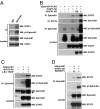
Fig. 1.
Fig. 1.
ephrinB1 associates with STAT3 in a phosphorylation-dependent manner. (A) IPs using control IgG or anti-ephrinB1 antibodies on mouse embryonic brain lysates (embryonic day 13.5) were immunoblotted with anti-STAT3 and anti-phosphotyrosine antibody. Lysates were analyzed directly by SDS/PAGE and Western blot analysis with indicated antibodies to reveal endogenous expression levels of STAT3 and ephrinB1, respectively. (B) Oocytes were left uninjected (−) or injected (+) with the indicated RNA(s). Oocyte lysates were immunoprecipitated with anti-FLAG (first and third rows) or anti-HA (second row) antibody, then immunoblotted with either anti-HA or anti-FLAG antibody to detect bound protein. The phosphorylation state was revealed by anti-phosphotyrosine antibody (third row). Oocyte lysates were analyzed directly by SDS/PAGE and Western blot analysis with anti-FLAG (fourth row), anti-HA (fifth row) antibody, and anti-FGFR1 antibody (sixth row). FGFR1KE, constitutively active Xenopus FGFR1 (FGFR1K562E); KD, kinase-dead Xenopus FGFR1 (FGFR1C289R/K420A); ephrinB1-FLAG, Flag-tagged Xenopus ephrinB1; STAT3-HA, HA-tagged Rat STAT3. (C) Extracts were prepared from HeLa cells transfected with ephrinB1 and STAT3 alone or along with an activated c-Src (Y529F) and were subjected to IP and immunoblot analysis with indicated antibodies. (D) Extracts were prepared from HeLa cells transfected with ephrinB1 and STAT3, were treated with control Fc or EphB2 ectodomain-Fc fusion, and were subjected to IP and immunoblot analysis with indicated antibodies.
Fig. 2.
Fig. 2.
ephrinB1 specifically recruits STAT3, resulting in tyrosine phosphorylation of STAT3 at position 705. (A) ephrinB1 RNA was injected into oocytes with STAT1-HA, STAT3-HA, and STAT5b-HA RNAs in the presence of FGFR1 KE or FGFR1 KD RNA. Oocyte lysates were immunoprecipitated with anti-ephrinB1 (first and third rows) or anti-HA (second row) antibody and then were immunoblotted with anti-HA to detect bound protein. The phosphorylation state was revealed by anti-phosphotyrosine antibody (third row). Oocyte lysates were analyzed directly by SDS/PAGE and Western blot analysis with anti-ephrinB1 (fourth row), anti-HA (fifth row) antibody, and anti-FGFR1 antibody (sixth row). (B and C) Oocytes were left uninjected (−) or injected (+) with the indicated RNA(s). Lysates were analyzed directly by SDS/PAGE and Western blot analysis with anti-pY705-STAT3 (B, first row), anti-phosphotyrosine (C, first row), anti-ephrinB1 (B and C, second row), anti-HA (B and C, third row), and anti-FGFR1 (B and C, fourth row) antibody.
Fig. 3.
Fig. 3.
Both Tyr 294 and Tyr 310 of ephrinB1 contribute to complex formation between ephrinB1 and STAT3. (A) A schematic representation of x-ephrinB1 cDNA harboring mutations at Tyr-294, Tyr-298, Tyr-305, and Tyr-310 in the conserved cytoplasmic domain. The C-terminal 33 aa of x-ephrinB1 are shown. Numbers indicate the position of the six conserved tyrosine residues, and the corresponding amino acids in murine ephrinB1 are shown in parentheses. (B) Xenopus oocytes were injected with the indicated combinations of x-ephrinB1 mutants, STAT3, and FGFR1 KE or FGFR1 KD. Lysates were immunoprecipitated using an anti-ephrinB1 (first, third, and fourth rows), anti-HA (second row) antibody. Coimmunoprecipitated STAT3 was detected using an anti-HA antibody (first row). Reblotting with anti-ephrinB1 antibody was performed (fourth row). Lysates were analyzed by Western blot analysis using an indicated antibody (fifth, sixth, and seventh rows). (C) Schematic diagrams of the structural domains of wild-type and mutant STAT3. Each domain is represented by boxes; mutation sites within specific functional domains are indicated by an asterisk. NT, N-terminal; CC, coiled-coil; DB, DNA binding; LN, linker; SH2, Src homology2; TA, transcriptional activation. (D) Xenopus oocytes were injected with RNAs encoding x-ephrinB1 and each STAT3 mutant in the presence of xFGFR1 KE or xFGFR1 KD. Lysates were immunoprecipitated with anti-ephrinB1 (first and third rows) and anti-HA antibody (second row), transferred, and immunoblotted with anti-HA and anti-phosphotyrosine antibody (second and third row). Lysates were analyzed directly by SDS/PAGE and Western blot analysis with anti-ephrinB1 (fourth row), anti-HA (fifth row), and anti-FGFR1 (sixth row) antibodies, respectively.
Fig. 4.
Fig. 4.
ephrinB1 increases STAT3 transcriptional activity through the induction of STAT3 phosphorylation. (A) COS-1 cells were transfected with ephrinB1 or a STAT3 binding mutant of ephrinB1 (Y294/Y310F), STAT3 or a DN-STAT3 mutant (Y705F), FGFR1 KE, pM67-tk-Luc, and pCMV-β-gal as indicated. The luciferase activity of each sample was normalized to β-gal activity. Data are represented as means ± SD and represent results from three independent experiments. (B) Protein extracts from A were directly assayed by Western blot analysis with antibodies to pY705-STAT3, ephrinB1, pY324-ephrinB1, and HA. (C) COS-1 cells were transfected with ephrinB1 or a STAT3-binding mutant of ephrinB1 (Y294/Y310F), STAT3 or a DN-STAT3 mutant (Y705F), pM67-tk-Luc, and pCMV-β-gal and treated with control Fc or EphB2-Fc as indicated. The luciferase activity of each sample was normalized to β-gal activity. Data are represented as means ± SD and represent results from three independent experiments. (D) Protein extracts from C were directly assayed by Western blot analysis with antibodies to pY705-STAT3, ephrinB1, and HA. (E) Neuroepithelial cells were treated with control Fc, EphB2-Fc, or FGF-2, and immunofluorescence was performed using the antibodies to pY705-STAT3 and DAPI staining of nuclei.
Fig. 5.
Fig. 5.
DN-Jak2 specifically inhibits ephrinB1-mediated STAT3 transcriptional activation. (A) ephrinB1, Jak2, and FGFR1 KE RNAs are coinjected into Xenopus oocytes as indicated. Lysates were immunoprecipitated with antibodies to ephrinB1 (first, second, and third rows) and subjected to Western blot analysis with indicated antibodies. Oocyte lysates were analyzed by SDS/PAGE and Western blotting with anti-ephrinB1 (fourth row), anti-HA (fifth row), and anti-FGFR1 (sixth row) antibody, respectively. (B) COS-1 cells were transiently transfected with ephrinB1, STAT3, FGFR1 KE, and DN-Jak1 or -2 or with DN-Tyk2, pM67-tk-Luc, and pCMV-β-gal as indicated. Luciferase activity was assayed as in Fig. 4A. (C) Protein extracts from B were directly assayed by Western blot analysis with antibodies to pY705-STAT3, Jak1, Tyk2, Jak2, ephrinB1, and HA, respectively.

References

    1. Palmer A, Klein R. Genes Dev. 2003;17:1429–1450. - PubMed
    1. Flanagan JG, Vanderhaeghen P. Annu Rev Neurosci. 1998;21:309–345. - PubMed
    1. Gale NW, Holland SJ, Valenzuela DM, Flenniken A, Pan L, Ryan TE, Henkemeyer M, Strebhardt K, Hirai H, Wilkinson DG, et al. Neuron. 1996;17:9–19. - PubMed
    1. Cowan CA, Henkemeyer M. Nature. 2001;413:174–179. - PubMed
    1. Lu Q, Sun EE, Klein RS, Flanagan JG. Cell. 2001;105:69–79. - PubMed

Publication types

MeSH terms