. 2007 Oct 30;104(44):17406-11.
doi: 10.1073/pnas.0703446104. Epub 2007 Oct 22.

High-resolution imaging of the dynamic tumor cell vascular interface in transparent zebrafish

Affiliations

High-resolution imaging of the dynamic tumor cell vascular interface in transparent zebrafish

Konstantin Stoletov et al. Proc Natl Acad Sci U S A. .

Abstract

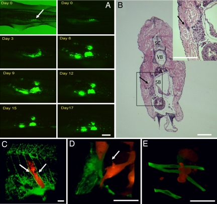
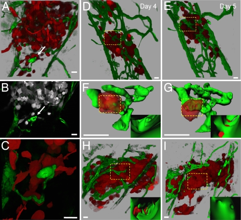
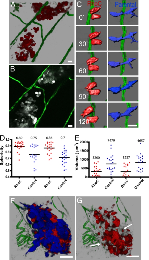
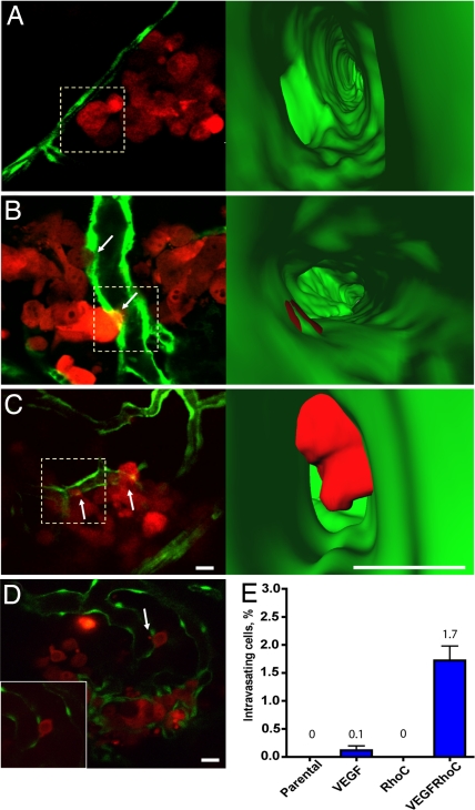
Cell metastasis is a highly dynamic process that occurs in multiple steps. Understanding this process has been limited by the inability to visualize tumor cell behavior in real time by using animal models. Here, we employ translucent zebrafish and high-resolution confocal microscopy to study how human cancer cells invade in tissues, induce angiogenesis, and interact with newly formed vessels. We use this system to study how the human metastatic gene RhoC promotes the initial steps of metastasis. We find that RhoC expression induces a primitive amoeboid-like cell invasion characterized by the formation of dynamic membrane protrusions and blebs. Surprisingly, these structures penetrate the blood vessel wall exclusively at sites of vascular remodeling and not at regions of existing intact vessels. This process requires tumor cells to secrete VEGF, which induces vascular openings, which in turn, serve as portholes allowing access of RhoC-expressing cells to the blood system. Our results support a model in which the early steps in intravasation and metastasis require two independent events: (i) dynamic regulation of the actin/myosin cytoskeleton within the tumor cell to form protrusive structures and (ii) vascular permeablization and vessel remodeling. The integration of zebrafish transgenic technology with human cancer biology may aid in the development of cancer models that target specific organs, tissues, or cell types within the tumors. Zebrafish could also provide a cost-effective means for the rapid development of therapeutic agents directed at blocking human cancer progression and tumor-induced angiogenesis.


Conflict of interest statement

The authors declare no conflict of interest.

Figures

Fig. 1.
Fig. 1.
Human tumor cells form microtumors in the body wall of zebrafish. (A) MDA-435 control cells expressing GFP were injected into the peritoneal cavity of zebrafish and imaged daily for 17 consecutive days with a fluorescence stereomicroscope. The image is representative of >100 injected animals. Arrow shows the injection site. (B) H&E stain of MDA-435 control cells 5 days after injection (dpi) in the body wall (cross-section). Tumor cells can be seen attached to the body wall surface (arrow). (Inset) High-magnification image of the box denoted in B. The arrow points to a group of cells invading into the body wall. SC, spinal cord; VB, vertebrae; SB, swim/air bladder. (C) Three-dimensional reconstruction of a microscopic MDA DsRed tumor developing in the body wall between the intersegmental vessels (arrows) of Tg(fli1:EGFP) fish, 4 dpi. (D) Three-dimensional reconstruction of single invading MDA-435 control cell. (E) Three-dimensional reconstruction of single invading HT1080 cell. Color code: In A, Human tumor cells are green; in C–E, fish blood vessels and green, and human tumor cells are red. [Scale bars, 1 mm (A and B); 200 μm (C), and 20 μm (D and E).]
Fig. 2.
Fig. 2.
Visualization of tumor-induced angiogenesis and tumor cell–vascular interactions. (A) Three-dimensional reconstruction of MDA-435 cell microtumor in the body wall of Tg(fli1:egfp) zebrafish tissue, 5 dpi. Note the remodeling vessel (white arrows in A–C). (B) Single optical section (1 μm) of microtumor in A, (SI Movie 1 shows a series of optical sections through the tumor and remodeling vessels). (C) High magnification of invasive cells clustering around the remodeling vessel shown in A and B (SI Movie 2). (D and E) Three-dimensional reconstruction of MDA-435 tumor cells secreting human VEGF in the body wall of Tg(fli1:egfp) zebrafish at 4 (D) and 5 (E) dpi. Images were obtained from the same animal on consecutive days. (F and G) Three-dimensional reconstructions of digitally isolated tumor cells in contact with host vessels from D and E (dotted squares; SI Movies 3 and 4). (Insets) Three-dimensional reconstructions of the vessel interior at sites of vessel openings and tumor cell membrane integration. (H) Three-dimensional reconstruction of a MDA-435 microtumor secreting human VEGF at 4 dpi before (H) and after (I) treatment (24 h) with 5 μm of the VEGF receptor inhibitor SU5416 (SI Movies 5 and 6). (Insets) Inside vessel surface within the dotted squares. Color code: Fish blood vessels are green, and human tumor cells are red (or gray in B). (Scale bars, 20 μm.)
Fig. 3.
Fig. 3.
RhoC-induced human tumor cell morphology and invasion. (A) Three-dimensional reconstruction of MDA-RhoC cells invading the body wall of Tg(fli1:egfp) zebrafish at 5 dpi. (B) Single optical section (1 μm) of cells in A (SI Movie 7 shows all optical sections through the tumor). (C) Time-lapse analysis of control MDA-435 (blue) and MDA-RhoC (red) cells invading within the body wall of Tg(fli1:egfp) zebrafish. Images were acquired every 15 min and reconstructed in 3-D and then digitally highlighted to reveal surface morphology by using Imaris Contoursurface. (D and E) The distribution of sphericity (round vs. elongated) (D) and cell volume (E) for MDA-435 and MDA-RhoC cells injected individually (●) or together (▴) were measured as described in SI Text. MDA-RhoC cells had significantly higher sphericity and lower volume (P < 0.05, t test) than MDA-control cells when injected separately or together. Horizontal lines represent mean values displayed above the plots. (F) Three-dimensional reconstruction of MDA-435 (blue) and MDA-RhoC cells (red) coinjected at a 1:1 ratio at 3 dpi. (G) Three-dimensional reconstruction of F, but only MDA-RhoC cells (red channel) are shown. Arrows indicate membrane blebs shed into the surrounding tissue. Color code: Fish blood vessels are green; in B–G, MDA-RhoC cells are red (or gray in B), and MDA parental cells are blue. (Scale bars, 20 μm.)
Fig. 4.
Fig. 4.
RhoC cooperates with VEGF to enhance tumor cell intravasation. Single optical sections showing tumor cells interacting with the vessel surface (5 dpi). (A–C) MDA-435 expressing RhoC (A), VEGF (B), or RhoC and VEGF (C). (Right) Three-dimensional reconstructions of interior vessel surfaces at the tumor cell-vessel interface (dotted squares in Left). Arrows show vascular mimicry (B) or membrane protrusion into the vessel lumen (C). In C, note the large increase in the membrane protrusion inside the vessel lumen of the MDA-RhoC cells secreting VEGF (SI Movies 8–10). (D) Single optical section showing an MDA-RhoC VEGF membrane protrusion in the vessel lumen (arrow). (Inset) The same cell 5 min later. (E) Plot showing percent of intravasating cells for parental MDA-435 cells or MDA-435 cells expressing VEGF, RhoC, or RhoC and VEGF. MDA-RhoC cells that express VEGF had a significantly higher percentage of intravasating cells (P < 0.05, t test) than other cell types. Mean and SEM values are displayed above the plot. Colors code: Fish vasculature is green, and human tumor cells are red. [Scale bars, 20 (Left) or 10 μm (Right).]

Similar articles

Cited by

References

    1. Gupta PB, Mani S, Yang J, Hartwell K, Weinberg RA. Cold Spring Harb Symp Quant Biol. 2005;70:291–297. - PubMed
    1. Gupta G, Massaque J. Cell. 2006;127:679–695. - PubMed
    1. Condeelis J, Segall JE. Nat Rev Cancer. 2003;3:921–930. - PubMed
    1. Zon LI, Peterson RT. Nat Rev Drug Discov. 2005;4:35–44. - PubMed
    1. Berghmans S, Jette C, Langenau D, Hsu K, Stewart R, Look T, Kanki JP. BioTechniques. 2005;39:227–237. - PubMed

Publication types

Substances