

Kinetics of synaptic transmission at ribbon synapses of rods and cones
- PMID: 17955196
- PMCID: PMC2474471
- DOI: 10.1007/s12035-007-0019-9
Kinetics of synaptic transmission at ribbon synapses of rods and cones
Abstract
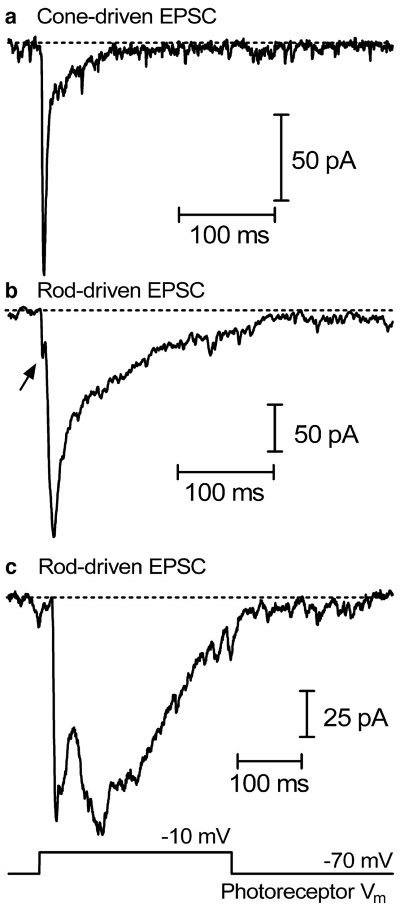
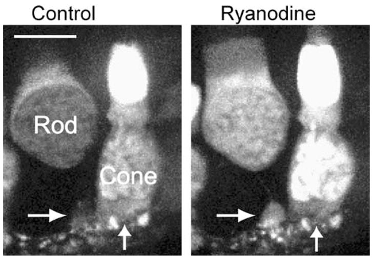
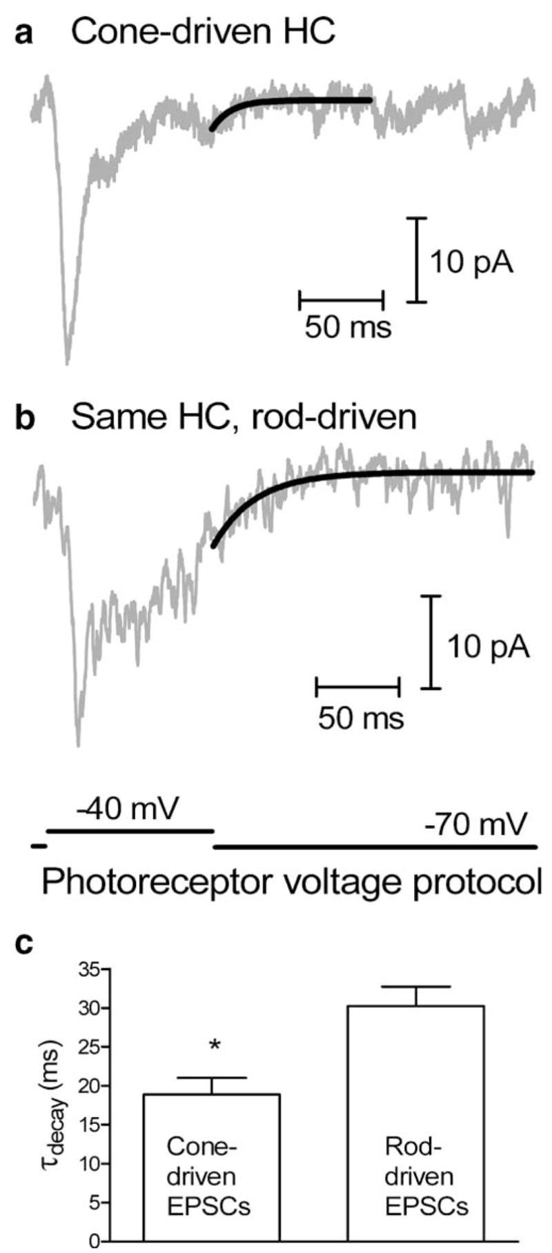
The ribbon synapse is a specialized structure that allows photoreceptors to sustain the continuous release of vesicles for hours upon hours and years upon years but also respond rapidly to momentary changes in illumination. Light responses of cones are faster than those of rods and, mirroring this difference, synaptic transmission from cones is also faster than transmission from rods. This review evaluates the various factors that regulate synaptic kinetics and contribute to kinetic differences between rod and cone synapses. Presynaptically, the release of glutamate-laden synaptic vesicles is regulated by properties of the synaptic proteins involved in exocytosis, influx of calcium through calcium channels, calcium release from intracellular stores, diffusion of calcium to the release site, calcium buffering, and extrusion of calcium from the cytoplasm. The rate of vesicle replenishment also limits the ability of the synapse to follow changes in release. Post-synaptic factors include properties of glutamate receptors, dynamics of glutamate diffusion through the cleft, and glutamate uptake by glutamate transporters. Thus, multiple synaptic mechanisms help to shape the responses of second-order horizontal and bipolar cells.
Figures
Similar articles
-
Mechanisms, pools, and sites of spontaneous vesicle release at synapses of rod and cone photoreceptors.Eur J Neurosci. 2016 Aug;44(3):2015-27. doi: 10.1111/ejn.13288. Epub 2016 Jun 22. Eur J Neurosci. 2016. PMID: 27255664 Free PMC article.
-
Calmodulin enhances ribbon replenishment and shapes filtering of synaptic transmission by cone photoreceptors.J Gen Physiol. 2014 Nov;144(5):357-78. doi: 10.1085/jgp.201411229. Epub 2014 Oct 13. J Gen Physiol. 2014. PMID: 25311636 Free PMC article.
-
Paired-pulse depression at photoreceptor synapses.J Neurosci. 2006 Mar 1;26(9):2555-63. doi: 10.1523/JNEUROSCI.3667-05.2006. J Neurosci. 2006. PMID: 16510733 Free PMC article.
-
Transmission at rod and cone ribbon synapses in the retina.Pflugers Arch. 2021 Sep;473(9):1469-1491. doi: 10.1007/s00424-021-02548-9. Epub 2021 Mar 29. Pflugers Arch. 2021. PMID: 33779813 Free PMC article. Review.
-
Molecular Components of Vesicle Cycling at the Rod Photoreceptor Ribbon Synapse.Adv Exp Med Biol. 2025;1468:325-330. doi: 10.1007/978-3-031-76550-6_54. Adv Exp Med Biol. 2025. PMID: 39930217 Review.
Cited by
-
Endogenous calcium buffering at photoreceptor synaptic terminals in salamander retina.Synapse. 2014 Nov;68(11):518-28. doi: 10.1002/syn.21768. Epub 2014 Jul 30. Synapse. 2014. PMID: 25049035 Free PMC article.
-
Differential regulation of cone calcium signals by different horizontal cell feedback mechanisms in the mouse retina.J Neurosci. 2014 Aug 27;34(35):11826-43. doi: 10.1523/JNEUROSCI.0272-14.2014. J Neurosci. 2014. PMID: 25164677 Free PMC article.
-
Overexpression of guanylate cyclase activating protein 2 in rod photoreceptors in vivo leads to morphological changes at the synaptic ribbon.PLoS One. 2012;7(8):e42994. doi: 10.1371/journal.pone.0042994. Epub 2012 Aug 13. PLoS One. 2012. PMID: 22912773 Free PMC article.
-
Tracking quantum dot-tagged calcium channels at vertebrate photoreceptor synapses: retinal slices and dissociated cells.Curr Protoc Neurosci. 2013 Jan;Chapter 2:Unit 2.18. doi: 10.1002/0471142301.ns0218s62. Curr Protoc Neurosci. 2013. PMID: 23315944 Free PMC article.
-
Electrically-evoked responses for retinal prostheses are differentially altered depending on ganglion cell types in outer retinal neurodegeneration caused by Crb1 gene mutation.Front Cell Neurosci. 2023 Feb 6;17:1115703. doi: 10.3389/fncel.2023.1115703. eCollection 2023. Front Cell Neurosci. 2023. PMID: 36814867 Free PMC article.
References
-
- Arshavsky VY, Lamb TD, Pugh EN., Jr G proteins and phototransduction. Annu Rev Physiol. 2002;64:153–187. - PubMed
-
- Burns ME, Baylor DA. Activation, deactivation, and adaptation in vertebrate photoreceptor cells. Annu Rev Neurosci. 2001;24:779–805. - PubMed
-
- Barnes S. After transduction: response shaping and control of transmission by ion channels of the photoreceptor inner segments. Neuroscience. 1994;58:447–459. - PubMed
-
- Sterling P, Matthews G. Structure and function of ribbon synapses. Trends Neurosci. 2005;28:20–29. - PubMed
Publication types
MeSH terms
Substances
Grants and funding
LinkOut - more resources
Full Text Sources
