. 2007 Oct 19;131(2):286-99.
doi: 10.1016/j.cell.2007.08.041.

Recycling of eukaryotic posttermination ribosomal complexes

Affiliations

Recycling of eukaryotic posttermination ribosomal complexes

Andrey V Pisarev et al. Cell. .

Abstract

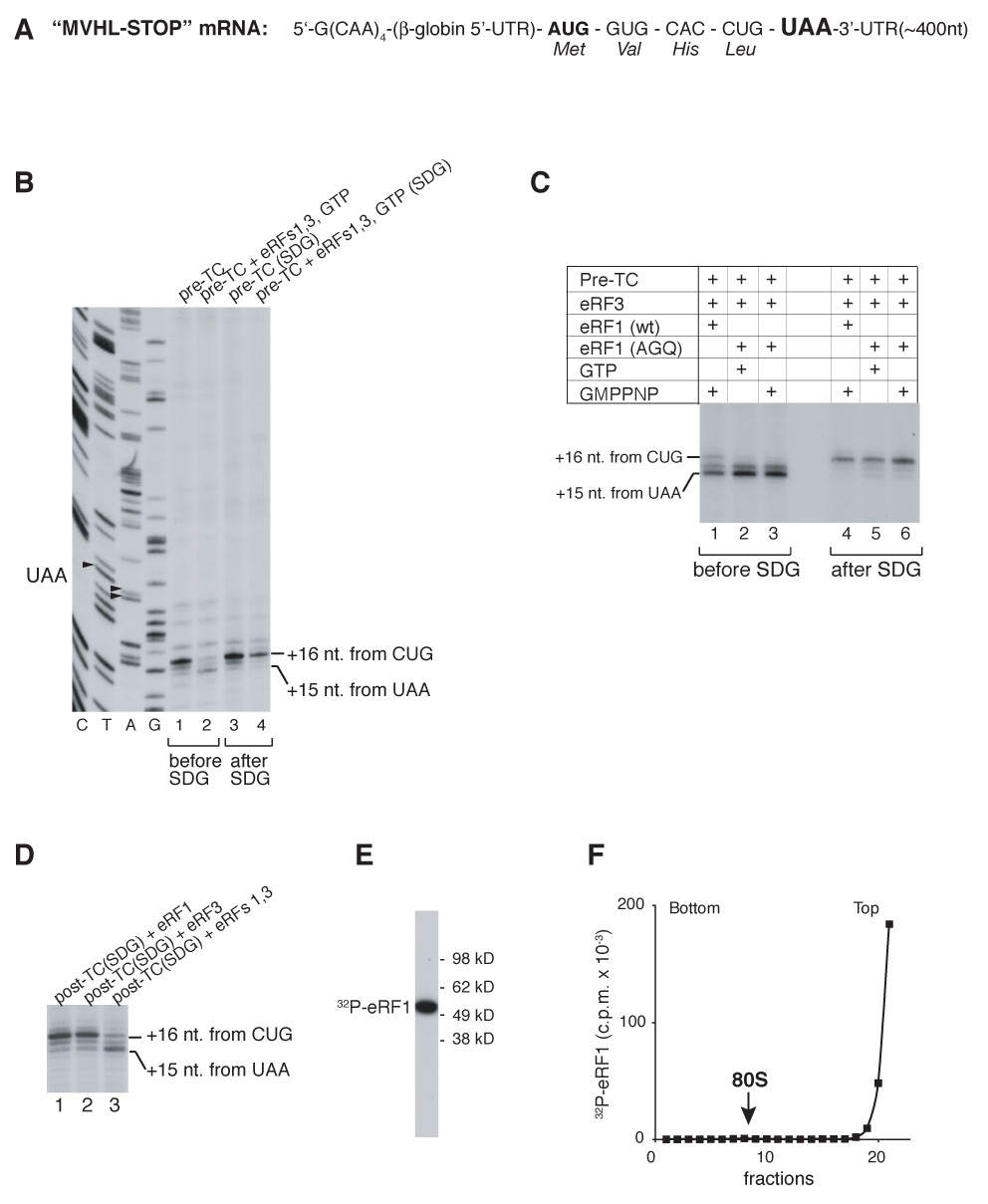
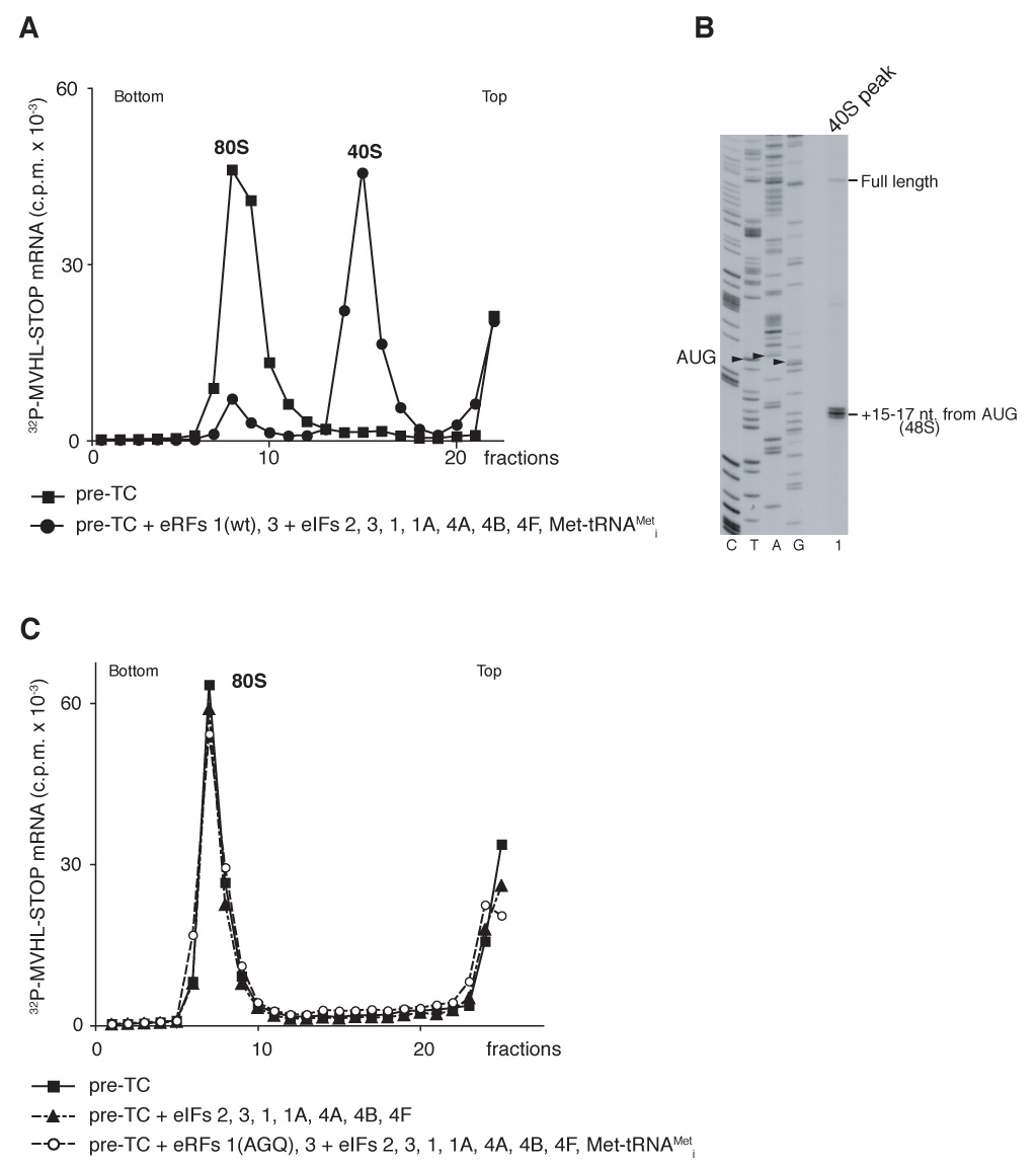
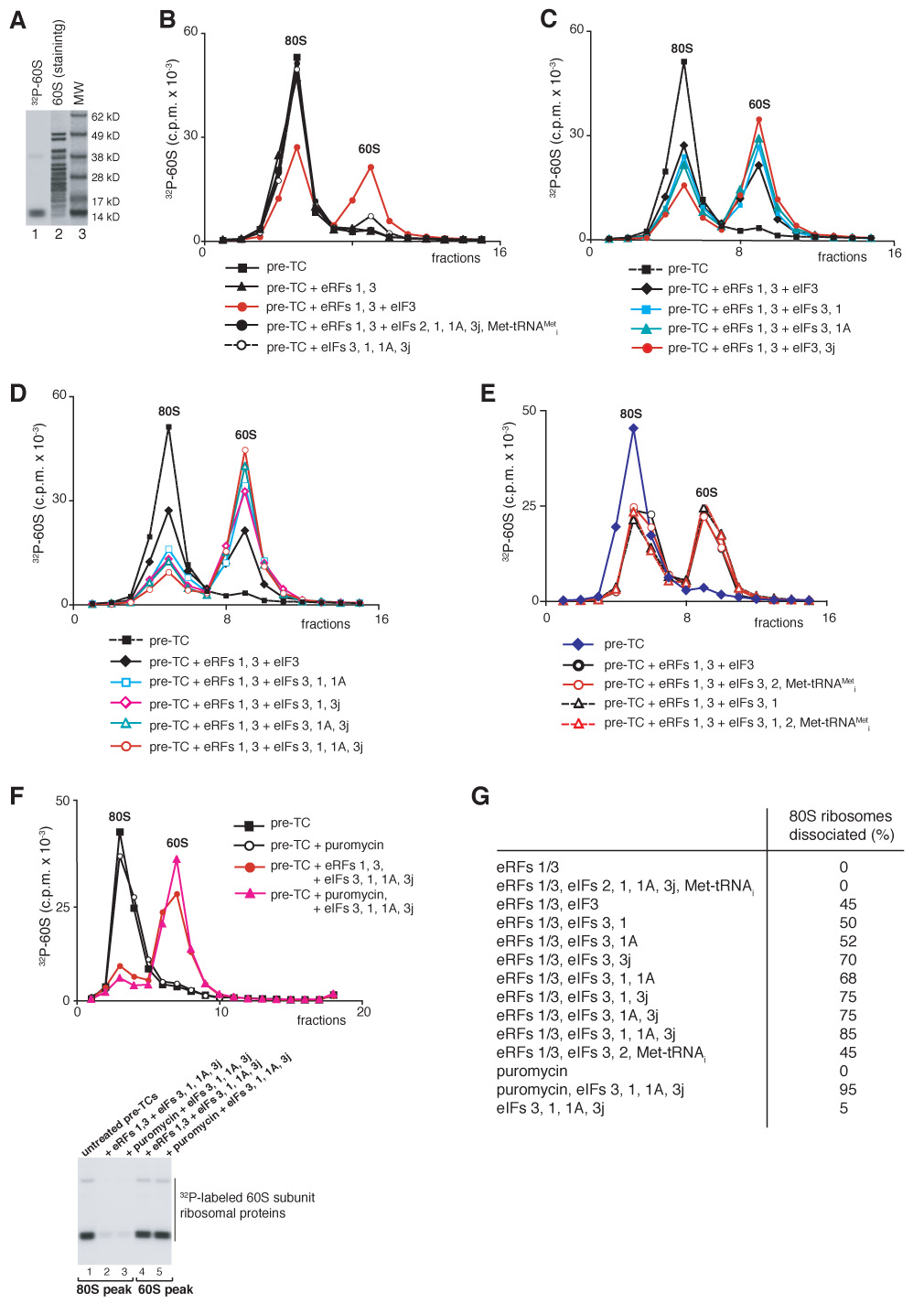
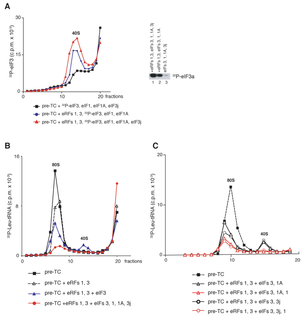
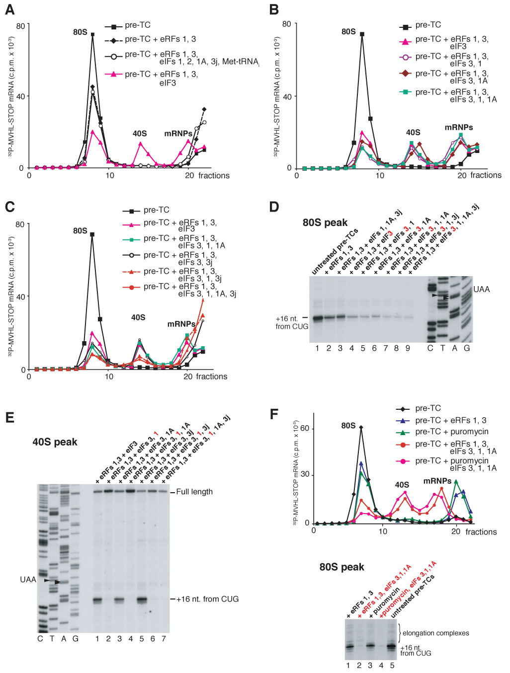
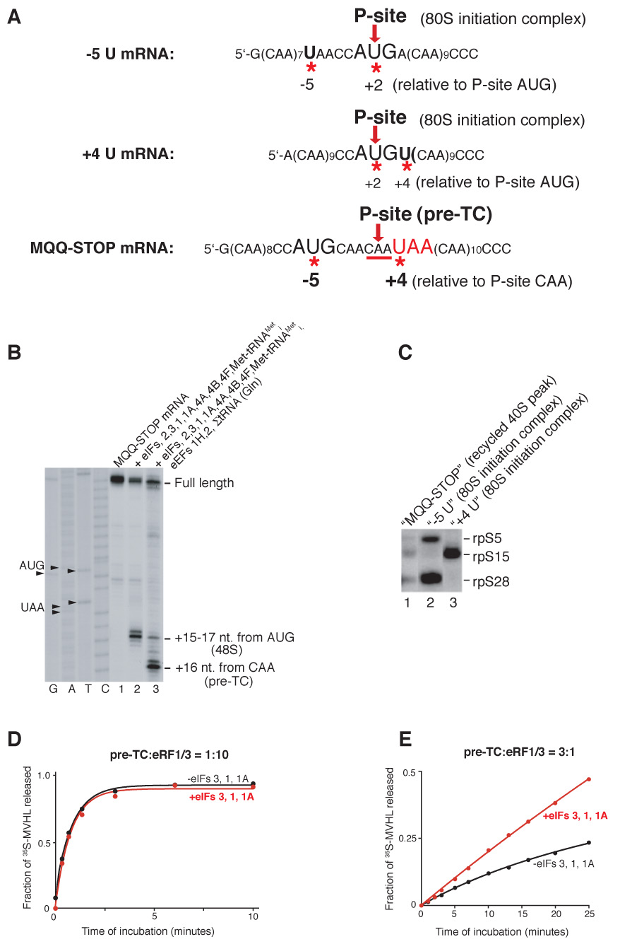
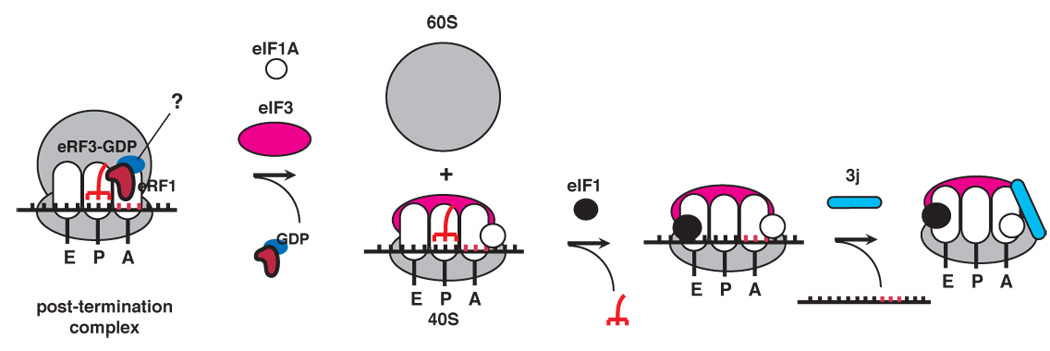
After translational termination, mRNA and P site deacylated tRNA remain associated with ribosomes in posttermination complexes (post-TCs), which must therefore be recycled by releasing mRNA and deacylated tRNA and by dissociating ribosomes into subunits. Recycling of bacterial post-TCs requires elongation factor EF-G and a ribosome recycling factor RRF. Eukaryotes do not encode a RRF homolog, and their mechanism of ribosomal recycling is unknown. We investigated eukaryotic recycling using post-TCs assembled on a model mRNA encoding a tetrapeptide followed by a UAA stop codon and report that initiation factors eIF3, eIF1, eIF1A, and eIF3j, a loosely associated subunit of eIF3, can promote recycling of eukaryotic post-TCs. eIF3 is the principal factor that promotes splitting of posttermination ribosomes into 60S subunits and tRNA- and mRNA-bound 40S subunits. Its activity is enhanced by eIFs 3j, 1, and 1A. eIF1 also mediates release of P site tRNA, whereas eIF3j ensures subsequent dissociation of mRNA.


Figures

Figure 1
Figure 1. Association of release factors with post-TCs
(A) Structure of MVHL-STOP mRNA. (B–D) Toe-printing analysis of ribosomal complexes obtained by incubating pre-TCs with eRFs, GTP and GMPPNP in different combinations, before and after sucrose gradient centrifugation, “SDG” (B, C) and of post-TCs, obtained by incubating pre-TCs with eRF1/eRF3/GTP, subjecting them to sucrose gradient centrifugation, and incubating them again with eRFs, as indicated (D). Lanes C, T, A, G depict cDNA sequences corresponding to MVHL-STOP mRNA. The positions of toe-prints that correspond to ribosomal complexes are indicated. (E) Autoradiograph of [32P]eRF1 phosphorylated by cAMP-dependent kinase. MW markers are indicated. (F) Association of [32P]eRF1 with post-TCs assayed by sucrose gradient centrifugation. The position of 80S ribosomes is indicated.
Figure 2
Figure 2. Recycling of post-TCs and assembly of 48S initiation complexes on recycled mRNA promoted by initiation factors
(A, C) Ribosomal association of [32P]MVHL-STOP mRNA after incubation of pre-TCs with eRFs and eIFs, as indicated, assayed by sucrose gradient centrifugation. The positions of 40S subunits and 80S ribosomes are indicated. (B) Toe-printing analysis of the 40S-containing fraction (panel A, circles) obtained after incubating pre-TCs with eRFs and eIFs. The positions of full-length cDNA and of toe-prints that correspond to 48S complexes are indicated. Lanes C, T, A, G depict cDNA sequences corresponding to MVHL-STOP mRNA.
Figure 3
Figure 3. Dissociation of post-TCs into subunits
(A) Coomassie staining of 60S subunit proteins (lane 2) and autoradiography of [32P]60S subunits phosphorylated by CKII (lane 1). MW markers are indicated. (B–F) Dissociation of pre-TCs, assembled on MVHL-STOP mRNA with [32P]60S subunits, after incubation with eRFs, eIFs and puromycin, as indicated, assayed after sucrose gradient centrifugation by Cerenkov counting and Pisarev, gel electrophoresis (F, right panel). The positions of 60S subunits and 80S ribosomes are indicated. (G) Summary of dissociation of post-termination ribosomes into subunits by different combination of eIFs (panels B–F).
Figure 4
Figure 4. Association of eIF3 with recycled 40S subunits and dissociation of P site deacylated tRNA
(A) Ribosomal association of [32P]eIF3 after incubation of pre-TCs assembled on MVHL-STOP mRNA with different combination of eRFs and eIFs, as indicated, assayed after sucrose gradient centrifugation by Cerenkov counting and gel electrophoresis (right panel). (B, C) Ribosomal association of [32P]tRNALeu after incubation of pre-TCs assembled in the presence of Leu-[32P]tRNALeu with eRFs and eIFs, as indicated, assayed after sucrose gradient centrifugation by Cerenkov counting. The positions of 40S subunits and 80S ribosomes are indicated.
Figure 5
Figure 5. Dissociation of mRNA from post-TCs
(A–C, F) Ribosomal association of [32P]MVHL-STOP mRNA after incubation of pre-TCs with eRFs, eIFs and puromycin, as indicated, assayed after sucrose gradient centrifugation by Cerenkov counting. The positions of 40S subunits, 80S ribosomes and mRNPs are indicated. (D,E, lower panel of F) Toe-printing analysis of 40S-containing fractions shown on panels A–C (E), and of 80S-containing fractions shown on panels A–C and F (D and F, respectively). The positions of full-length cDNA and of toe-prints that correspond to ribosomal complexes are indicated. Lanes C, T, A, G depict cDNA sequences corresponding to MVHL-STOP mRNA.
Figure 6
Figure 6. The position of mRNA on recycled 40S subunits and the influence of initiation factors on peptide release
(A) Sequences of −5U, +4U and MQQ-STOP mRNAs. The positions of Us (indicated by red asterisks) relative to the P-site codons (red arrows) in 80S initiation complexes or in pre-TCs are indicated. (B) Toe-printing analysis of 48S complexes and pre-TCs assembled on MQQ-STOP mRNA. The components of reaction mixtures are indicated. The position of ribosomal complexes are shown relative to the mRNA codon in the P site. Lanes C, T, A, G depict cDNA sequences corresponding to MQQ-STOP mRNA. (C) UV cross-linking of 32P-labeled −5U, +4U and MQQ-STOP mRNAs containing 4-ThioU with ribosomal proteins in recycled 40S subunit-containing complexes or in 80S initiation complexes, as indicated, assayed by SDS-PAGE and autoradiography. (D, E) Kinetics of [35S]MVHL tetrapeptide release in the presence (red circles) and in the absence (black circles) of eIFs at different ratios of eRFs and pre-TCs.
Figure 7
Figure 7. A model for eukaryotic ribosomal recycling

References

    1. Alkalaeva EZ, Pisarev AV, Frolova LY, Kisselev LL, Pestova TV. In vitro reconstitution of eukaryotic translation reveals cooperativity between release factors eRF1 and eRF3. Cell. 2006;125:1125–1136. - PubMed
    1. Carter AP, Clemons WM, Jr, Brodersen DE, Morgan-Warren RJ, Hartsch T, Wimberly BT, Ramakrishnan V. Crystal structure of an initiation factor bound to the 30S ribosomal subunit. Science. 2001;291:498–501. - PubMed
    1. Dallas A, Noller HF. Interaction of translation initiation factor 3 with the 30S ribosomal subunit. Mol. Cell. 2001;8:855–864. - PubMed
    1. Demeshkina N, Hirokawa G, Kaji A, Kaji H. Novel activity of eukaryotic translocase, eEF2: dissociation of the 80S ribosome into subunits with ATP but not with GTP. Nucleic Acids Res. 2007;35:4597–4607. - PMC - PubMed
    1. Fraser CS, Berry KE, Hershey JWB, Doudna JA. eIF3j is located in the decoding center of the human 40S ribosomal subunit. Mol. Cell. 2007;26:811–819. - PubMed

Publication types

MeSH terms

LinkOut - more resources