. 2007 Oct 19;131(2):337-50.
doi: 10.1016/j.cell.2007.08.030.

Cordon-bleu is an actin nucleation factor and controls neuronal morphology

Affiliations

Cordon-bleu is an actin nucleation factor and controls neuronal morphology

Rashmi Ahuja et al. Cell. .

Abstract

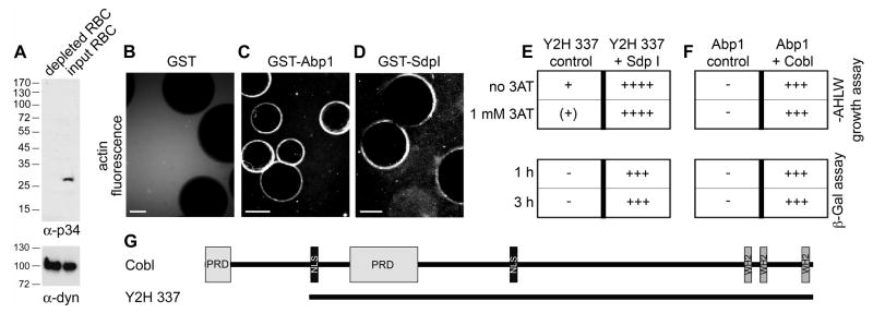
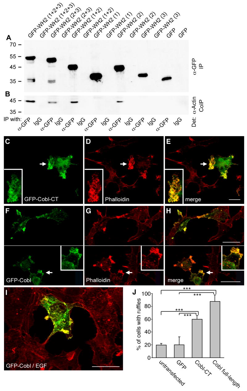
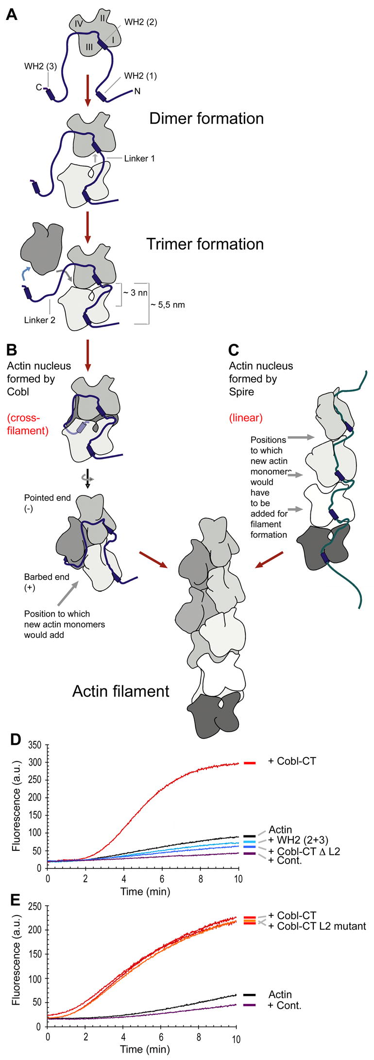
Despite the wealth of different actin structures formed, only two actin nucleation factors are well established in vertebrates: the Arp2/3 complex and formins. Here, we describe a further nucleator, cordon-bleu (Cobl). Cobl is a brain-enriched protein using three Wiskott-Aldrich syndrome protein homology 2 (WH2) domains for actin binding. Cobl promotes nonbundled, unbranched filaments. Filament formation relies on barbed-end growth and requires all three Cobl WH2 domains and the extended linker L2. We suggest that the nucleation power of Cobl is based on the assembly of three actin monomers in cross-filament orientation. Cobl localizes to sites of high actin dynamics and modulates cell morphology. In neurons, induction of both neurites and neurite branching is dramatically increased by Cobl expression-effects that critically depend on Cobl's actin nucleation ability. Correspondingly, Cobl depletion results in decreased dendritic arborization. Thus, Cobl is an actin nucleator controlling neuronal morphology and development.


Figures

Figure 1
Figure 1. Identification of Cobl
(A) Rat brain cytosol was depleted for the Arp2/3 complex, as demonstrated by anti-p34 immunoblotting and using the anti-dynamin signal as loading control. (B-D) Both immobilized GST-Abp1 (C) and syndapin I (D) but not GST alone (B) were still able to elicit fluorescent F-actin halos in Arp2/3 complex-depleted cytosol endowed with Alexa Fluor™ 568 labeled G-actin. Scale bars, 25 μm. (E and F) Directed yeast-2-hybrid analyses of the isolated clone 337 (Y2H 337) as a prey with syndapin I as a bait (E) as well as Y2H 337 as a bait and Abp1 as a prey (F), respectively, caused strong activity of independent reporter gene systems. (G) Domain structure of murine Cobl (gi32251014) and of clone Y2H 337. Domain abbreviations, PRD, proline-rich domain, NLS, putative nuclear localization signal, WH2, WASP-homology 2.
Figure 2
Figure 2. The individual Cobl WH2 domains are highly related and sequester G-actin
(A) Sequence alignment of different WH2 motifs by Clustal W 1.83 with depicted position of the amphiphatic α-helix. Identical residues are shaded in dark grey; those with conservation or similarities in at least two of the Cobl WH2 domains are shaded in light grey. (B) Phylogenetic analysis of WH2 domains by Clustal W (1.83) (alignment see Figure S3). (C) Affinity purifications of actin from rat brain extracts with equal amounts of GST-Cobl WH2 domains. (D-G) Actin bead assay with Cobl GST-WH2 domains. Images after 5 minutes (WH2 (1) and WH2 (2) (D, E)) and 30 minutes of incubation (WH2 (3) (F)) show actin on the surface. Beads coated with GST alone did not accumulate any actin (G). Bar in F for D, E and F, 15 μm; bar in G, 10 μm. (H-J) In vitro reconstitutions of actin dynamics (pyrene-actin assays). Actin (actin, 4 μM; pyrene-actin, 0.4 μM) was polymerized in the presence of the indicated concentrations of GST-WH2 (1) (H), GST-WH2 (2) (I) and GST-WH2 (3) (J). WH2 (1) and (2) quench G-actin very effectively and thus prevent spontaneous actin filament formation. Insets show Coomassie stainings of the WH2 domains. (K-M) Determination of G-actin dissociation constants of WH2 (1) (K), WH2 (2) (L), and WH2 (3) (M) by measuring the fluorescence of NBD-labeled G-actin (0.4 μM) incubated with different concentrations of the WH2 domains.
Figure 3
Figure 3. Cobl WH2 domains bind actin in vivo and Cobl induces the formation of actin-rich ruffles in COS-7 cells
(A and B) Coimmunoprecipitation of endogenous actin from HEK293 cells with different GFP-Cobl constructs. Immunoblot analysis of immunoprecipitated GFP-fusion proteins (A) and of coimmunoprecipitated endogenous actin (B). (C-H) COS-7 cells transiently transfected with GFP-Cobl-CT (C-E) and GFP-Cobl full-length (F-H) exhibit ruffles enriched for the Cobl proteins (C and F; arrows) and F-actin detected by phalloidin staining and confocal microscopy (D and G; arrows). Insets are enlargements of the ruffles marked by arrows. (I) EGF-treated cells (15 min, final concentration 100 ng/ml) show a colocalization of Cobl full-length (green) and F-actin (red) in ruffles, too. (J) Quantification of the percentage of cells with ruffle induction (n > 300 each; 3-6 independent assays). Data are represented as mean ± SEM (p < 0.001, ***). Bar, 12 μm.
Figure 4
Figure 4. Cobl-CT promotes the assembly of actin filaments
(A, B and D) In vitro reconstitutions of actin nucleation and polymerization. (A) Cobl-CT nucleates actin filament assembly in a dose-dependent manner (2 μM actin) and shortens the lag phase when compared to actin alone and to addition of Flag-GFP (Cont.), respectively. Actin polymerization induced by 20 nM Arp2/3 complex and 60 nM N-WASP WA is shown for comparison. (B) At higher Cobl-CT concentrations, a dose-dependent decrease of Cobl-induced actin filament formation is observed. (C) Relative rate of actin assembly (maximal slope measured during polymerization) for different concentrations of Cobl-CT plotted on a logarithmic (black) and a linear scale (green). (D) Comparison of Cobl-CT to WH2 (1+2) and WH2 (2+3) shows that all three WH2 domains are required. (E-H) Visual evaluation of actin structures produced by Cobl-CT and Arp2/3. Filaments produced by 40 nM of Cobl-CT are long and mostly unbranched (E), whereas those produced by 25 nM of Arp2/3 complex and 100 nM N-WASP WA are often branched (F). Bar, 10 μm. (G) Quantitative analysis of the percentage of filaments appearing branched. (H) Quantitative analysis of filament lengths produced by Arp2/3 plus WA and Cobl-CT, respectively (n= 150 filaments each).
Figure 5
Figure 5. Cobl affects the morphology of primary hippocampal neurons
(A) Characterization of rabbit anti-Cobl antibodies in immunoblot experiments. (B) Western blot analysis of postnuclear supernatants of various rat tissues (100 μg protein each) probed with antibodies against Cobl. (C-E) Colocalization (E, merge) of endogenous Cobl detected by anti-Cobl antibodies (C) with phalloidin-stained F-actin (D) in growth cones of primary hippocampal neurons at DIV2 analyzed by confocal microscopy. Insets represent high magnifications of the axonal growth cone (arrowheads). Lower panels show a cell with more F-actin-rich dendritic growth cones (arrows). Bar in E, 20 μm. (F-N) Morphological analyses of Cobl functions in developing primary hippocampal neurons transfected at DIV1. 48 h later, neurons expressing GFP-Cobl-CT (F), GFP (H), GFP-WH2 (2+3) and GFP-WH2 (1+2), respectively, were costained for MAP2 (G and I) and evaluated for length of axon (J; n= 80 cells each), branching points per axon (K; n= 55), branching points of the dendritic arbor (L; n= 55), dendrites per cell (M; n= 55) and dendrite length (N; n= 55). Bar in I, 20 μm. (O-T) Morphological analyses of Cobl functions in hippocampal neurons transfected at DIV5 with GFP-Cobl full-length (O), GFP control (Q), GFP-WH2 (1+2) and GFP-WH2 (2+3) (S, T). Neuronal morphology was analyzed based on MAP2 immunostainings (P, R) 48 later. Quantitative analyses (n= 60) confirmed that Cobl-CT and especially Cobl full-length expression led to an increase of branching points (S) as well of neurites (T) compared to WH2 (2+3), WH2 (1+2) and GFP expressing neurons. Bar in O, 15 μm. (U) RNAi-mediated knock down of Cobl shown by anti-GFP immunoblotting of extracts from COS-7 cells transfected with bicistronic derivatives of the RNAi tools additionally encoding for GFP-Cobl(1-408). (V-Y) Morphology of Cobl-depleted neurons (V) transfected at DIV5 versus control (X), as detected by cell fillers (GFP, RFP; V, X) and MAP2 labeling (W, Y). Bar in Y, 20 μm. Quantitative analyses show that RNAi-mediated knock down of Cobl leads to fewer neurites (Ä) and a loss of branching (Z) when compared to controls, such as a non-functional and an unrelated RNAi construct, empty RNAi vector pRNAT, GFP, RFP (n=80 each) as well as rescue experiments with Cobl mutants insensitive for RNAi#1 and #2 (n=50 each). Data are represented as mean ± SEM (p < 0.05, *; p < 0.01, **; p < 0.001, ***).
Figure 6
Figure 6. Cobl-CT nucleates barbed-end actin polymerization in a mechanism distinct from that of the Arp2/3 complex
(A) GFP-Cobl-CT but not WH2 (1+2), WH2 (2+3) and the linker L2 specifically associates with F-actin, as shown by cosedimentation assays with 1 μM protein and 20 μM actin. Supernatant (S) and pellet (P) fractions were immunoblotted with anti-GFP (upper panel) and detected by Coomassie staining (actin), respectively (middle panel). The lower panel shows control reactions without actin. (B; C) Cytochalasin D (CD) completely inhibits Cobl-CT-induced actin assembly. Actin (2 μM; 0.2 μM pyrene-actin) was assembled in the presence and absence of 20 nM CD and Cobl-CT (10 nM). 100 nM Cytochalasin B (CB) exhibited similar effects. (D) Cobl and the Arp2/3 complex (40 nM each) promote filament assembly independently from each other. (E-G) Cobl-CT does not interfere with actin filament disassembly from the pointed end but slows disassembly at concentrations below the critical concentration of barbed ends. Preformed pyrene-labeled actin filaments (2 μM actin; 0.2 μM pyrene-actin) were diluted to a final concentration of 0.1 μM (E) and 0.05 μM (F), respectively. (G) Quantitative determinations of depolymerization rates, i.e. of slopes of dilution-induced depolymerization - normalized to corresponding experiments with actin alone.
Figure 7
Figure 7. Working model of Cobl-induced actin nucleation by cross-filament assembly of an actin trimer
(A) Model of Cobl/actin complex formation. Throughout the figure, the N-terminal, amphiphatic α-helix of the Cobl WH2 domains was placed into the shear zone between actin subdomains 1 and 3, which are oriented towards the barbed end, based on crystal structures of other WH2-proteins (Hertzog et al., 2004; Irobi et al., 2004). (B) Proposed structure of a Cobl-induced actin nucleus (front view and rotated by 90°) and mechanism of filament formation (elongation). (C) Filament formation and elongation by addition of actin monomers to linear actin trimers proposed to be assembled by Spire (Quinlan et al., 2005). (D) Experimental test of the cross-filament nucleation hypothesis by deleting the extended linker L2, which allows for the third WH2 domain to reach the backside of a forming nucleus. Cobl-CT ΔL2 (40 nM) failed to induce actin nucleation. Results were indistinguishable from WH2 deletions and GFP control. (E) Cobl-CT with a mutated L2 sequence of similar length (orange) nucleates as effectively as wild-type Cobl-CT (two examples in red).

Comment in

References

    1. Blanchoin L, Amann KJ, Higgs HN, Marchand JB, Kaiser DA, Pollard TD. Direct observation of dendritic actin filament networks nucleated by Arp2/3 complex and WASP/Scar proteins. Nature. 2000;404:1007–11. - PubMed
    1. Braun A, Pinyol R, Koch D, Dahlhaus R, Fonarev P, Grant BD, Kessels MM, Qualmann B. EHD proteins associate with syndapin I and II and such interactions play a crucial role in endosomal recycling. Mol Biol Cell. 2005;16:3642–3658. - PMC - PubMed
    1. Carroll EA, Gerrelli D, Gasca S, Berg E, Beier DR, Copp AJ, Klingensmith J. Cordon-bleu is a conserved gene involved in neural tube formation. Dev Biol. 2003;262:16–31. - PubMed
    1. Chereau D, Kerff F, Graceffa P, Grabarek Z, Langsetmo K, Dominguez R. Actin-bound structures of WASP-homology domain 2 and the implications for filament assembly. Proc Natl Acad Sci U S A. 2005;102:16644–16649. - PMC - PubMed
    1. Co C, Wong DT, Gierke S, Chang V, Taunton J. Mechanism of actin network attachment to moving membranes: barbed end capture by N-WASP WH2 domains. Cell. 2007;128:901–913. - PMC - PubMed

Publication types