Comparative Study
. 2007 Nov;130(Pt 11):2800-15.
doi: 10.1093/brain/awm236.

Lesion genesis in a subset of patients with multiple sclerosis: a role for innate immunity?

Affiliations
Comparative Study

Lesion genesis in a subset of patients with multiple sclerosis: a role for innate immunity?

Christina Marik et al. Brain. 2007 Nov.

Abstract

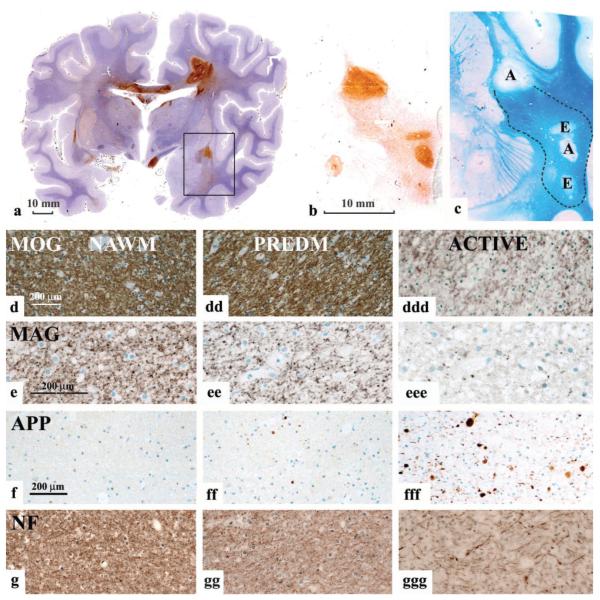
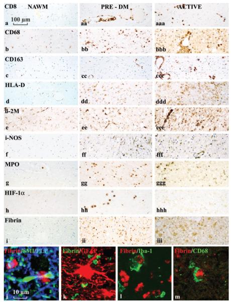
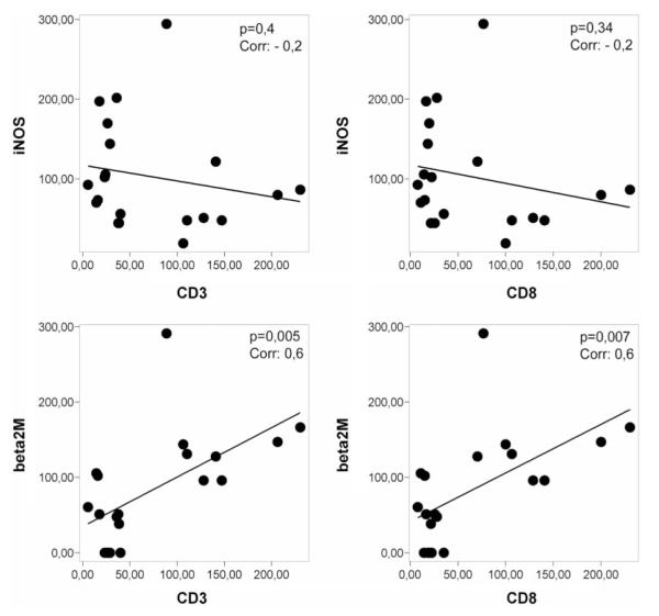
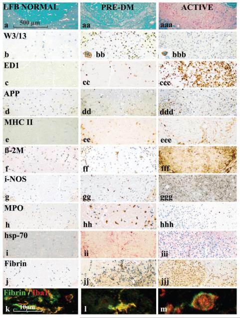
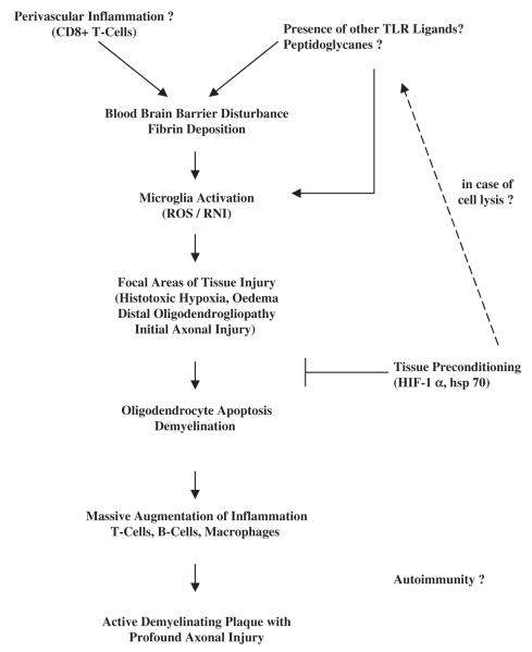
Lesions obtained early in the course of multiple sclerosis (MS) have been studied immunocytochemically, and compared with the early stages of the experimental lesion induced in rats by the intraspinal injection of lipopolysaccharide. Large hemispheric or double hemispheric sections were examined from patients who had died in the course of acute or early relapsing multiple sclerosis. In MS patients exhibiting hypoxia-like lesions [Pattern III; Lucchinetti et al. Ann Neurol (2000) 47: 707-17], focal areas in the white matter showed mild oedema, microglial activation and mild axonal injury in the absence of overt demyelination. In such lesions T-cell infiltration was mild and restricted to the perivascular space. Myeloperoxidase and the inducible form of nitric oxide synthase were expressed primarily by microglia, and the activated form of these cells was associated with extracellular deposition of precipitated fibrin. In addition, these lesions showed up-regulation of proteins involved in tissue preconditioning. When active demyelination started, lesions were associated with massive T-cell infiltration and microglia and macrophages expressed all activation markers studied. Similar tissue alterations were found in rats in the pre-demyelinating stage of lesions induced by the focal injection of bacterial lipopolysaccharide into the spinal white matter. We suggest that the areas of microglial activation represent an early stage of tissue injury, which precedes the formation of hypoxia-like demyelinated plaques. The findings indicate that mechanisms associated with innate immunity may play a role in the formation of hypoxia-like demyelinating lesions in MS.


Figures

Fig. 1
Fig. 1
Pattern III lesions. (a) Hemispheric brain section, labelled immunocytochemically for CD 68 (Case Pattern III C). There are multiple actively demyelinating lesions that show very dense immunoreactivity for CD68. In addition there are broad areas around the plaques, as well as areas independent from plaques that show weak reactivity for CD68 (areas of microglial activation). The rectangle indicates the area of the brain, shown in Fig. b and c. (b and c) Higher magnification of the area shown in the rectangle in (a). Serial sections are labelled immunocytochemically for CD 68 (b), and stained with Luxol fast blue (c). Areas with strong CD68 reactivity indicate areas of active demyelination (labelled with A in Fig. 1c). Areas with moderate CD68 expression show oedema in LFB-stained sections in the absence of active demyelination (E in Fig.1c). In addition there is a broad area of diffuse microglial activation (Fig.1b) between the active plaques, which shows a variable reduction in myelin density (lower part of the figures); this area is outlined in dashed lines in Fig. 1c. (d–g) Immunocytochemistry for myelin antigens (indicated), for APP and neurofilament (NF) in the normal appearing white matter (NAWM; d, e, f, g). Areas of diffuse microglial activation (‘pre demyelinating’: PREDM; dd, ee, ff, gg) and areas of actively demyelinating lesion (ddd, eee, fff, ggg) are shown. In comparison with the NAWM,‘pre-demyelinating’ lesions show similar MOG immunoreactivity, no demyelination and no macrophages with myelin degradation products, but some vacuoles, suggesting oedema. Immunoreactivity for MAG is profoundly reduced in ‘pre-demyelinating area’ in comparison to NAWM, and the labelling is in part fragmented into small granular structures. Some axons with immunoreactivity for amyloid precursor protein are seen in the ‘pre-demyelinating’ area, suggesting acute axonal injury, but there is no visible axonal loss in sections stained for neurofilament. In classical, active lesions there is a massive reduction in immunoreactivity for MOG, but there are still MOG-reactive fibres present. There are also many macrophages with MOG-reactive degradation products. In contrast, MAG reactivity is completely lost; numerous axons contain immunoreactivity for APP and the axonal density is clearly reduced in sections stained for neurofilament.
Fig. 2
Fig. 2
Inflammation and microglial activation in different stages of Pattern III lesions in comparison with that in the NAWM. This figure is organized into three columns, showing tissue from the NAWM,‘pre-demyelinating’ and actively demyelinating lesions respectively, in MS patients exhibiting Pattern III demyelination. The rows show the expression of CD8+ (MHC I-restricted T-cells (a), different macrophage and microglial activation markers (b–g), HIF-1α (h) and fibrin (i–m). T-cells in ‘pre-demyelinating’ lesions are restricted to the perivascular space (aa), while they diffusely disperse into the lesion in active plaques (aaa).‘Pre-demyelinating’ lesions show profound upregulation in microglia of CD68, HLA-D, iNOS and MPO, while in active lesions the most extensive expression is seen for CD68 and MHC Class I (β2M). HIF-1α is mainly expressed in ‘pre-demyelinating’ lesions. Immunocytochemistry for fibrin shows fibrin precipitates in ‘pre-demyelinating’ lesions, while in active lesions the immunoreactivity is diffuse, and also, in part, present within the cytoplasm of activated astrocytes. (j–m) Confocal double labelling for fibrin and other cellular markers (neurofilament and PLP in j; GFAP in k; AIF-1/Iba-1 in l and CD68 in m). Because there is no co-labelling, we conclude that the fibrin precipitates are present in the extracellular space, although they are closely attached to axonal spheroids (j) and to activated microglial cells (l and m).
Fig. 3
Fig. 3
Correlation between T-cell infiltration in lesion areas and expression of microglia activation antigens. There is a significant correlation between the number of CD3+ T-cells and the expression of MHC-class I in microglia within lesions. This is not the case for iNOS expression.
Fig. 4
Fig. 4
Inflammation and microglial activation in the LPS-induced demyelinating lesion during the pre-demyelinating phase and the phase of active demyelination. The figure is organized into three columns, showing respectively normal control tissue, and lesioned tissue during the pre-demyelinating period (1 to 5 days post-LPS injection) and the actively demyelinating period (12 days post-LPS injection). In comparison with normal spinal cord there is some reduction of myelin density (LFB staining) in the pre-demyelinating stage, primarily due to oedema, and profound loss of myelin staining in the active lesions (row a). W3/13, a marker for granulocytes and T-cells, shows profound granulocyte and T-cell infiltration of the tissue in the early pre-demyelinating stage (8 h and 1day after LPS injection), with moderateT-cell infiltration in active lesions (see also inserts). APP staining shows some axonal injury in the ‘pre-demyelinating’ stage and massive axonal damage when lesions actively demyelinate. The expression patterns of microglia/macrophage activation antigens is essentially similar to that seen in Pattern III MS lesions (Fig. 3), although the expression of these molecules is mainly found in cells with a macrophage phenotype (round cells) rather than a microglial appearance. Hsp 70 (visualized in red colour) as a marker for tissue preconditioning is mainly expressed during the pre-demyelinating stage. Immunocytochemistry for fibrin shows fibrin precipitates (dark-brown structures) in the pre-demyelinating stages, but diffuse reactivity in the active lesions. (k–m) Confocal double labelling of fibrin precipitates in the pre-demyelinating stage with the microglia/macrophage marker AIF-1/iba-1 shows precipitation of fibrin on the surface of microglial cells and macrophages.
Fig. 5
Fig. 5
Hypopthetical pathway of lesion formation in pattern III MS cases. TLR = toll-like receptors; ROS = reactive oxygen species; RNI = reactive nitrogen intermediates; HIF = hypoxia-inducible factor; hsp = heat shock protein.

Comment in

References

    1. Aboul-Enein F, Rauschka H, Kornel B, Stadelmann C, Stefferl A, Brück W, et al. Preferential loss of myelin associated glycoprotein reflects hypoxialike white matter damage in stroke and inflammatory brain diseases. J Neuropath Exp Neurol. 2003;62:25–33. - PubMed
    1. Adams RA, Bauer J, Flick MJ, Sikorski S, Nuriel T, Lassmann H, et al. The fibrin-derived γ377–395 peptide inhibits microglia activation and suppresses relapsing paralysis in central nervous system autoimmune disease. J Exp Med. 2007;204:571–82. - PMC - PubMed
    1. Akassoglou K, Adams RA, Bauer J, Mercado P, Tseveleki V, Lassmann H, et al. Fibrin depletion decreases inflammation and delays the onset of demyelination in a tumor necrosis factor transgenic mouse model for multiple sclerosis. Proc Natl Acad Sci USA. 2004;101:6698–703. - PMC - PubMed
    1. Altieri DC, Bader R, Mannucci PM, Edgington TS. Oligospecificity of the cellular adhesion receptor Mac-1 encompasses an inducible recognition specificity for fibrinogen. J Cell Biol. 1988;107:1893–900. - PMC - PubMed
    1. Babinski J. Recherches sur l’anatomie pathologique de la sclerose en plaque et etude comparative des diverses varietes de la scleroses de la moelle. Arch Physiol (Paris) 1885;5–6:186–207.

Publication types

MeSH terms