

Placental productions and expressions of soluble endoglin, soluble fms-like tyrosine kinase receptor-1, and placental growth factor in normal and preeclamptic pregnancies
- PMID: 17956952
- PMCID: PMC2190747
- DOI: 10.1210/jc.2007-1550
Placental productions and expressions of soluble endoglin, soluble fms-like tyrosine kinase receptor-1, and placental growth factor in normal and preeclamptic pregnancies
Abstract
Context: Increased production of antiangiogenic factors soluble endoglin (sEng) and soluble fms-like tyrosine kinase receptor-1 (sFlt-1) by the placenta contributes to the pathophysiology in preeclampsia (PE).
Objective: Our objective was to determine the differences in endoglin (Eng), fms-like tyrosine kinase receptor-1 (Flt-1), and placental growth factor (PlGF) expressions between normal and PE placentas and sEng, sFlt-1, and PlGF production by trophoblast cells (TC) cultured under lowered oxygen conditions.
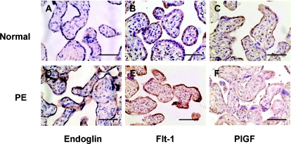
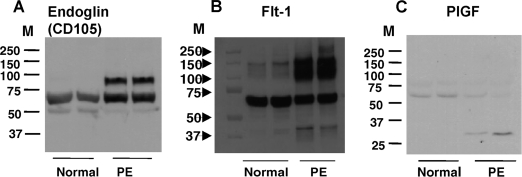
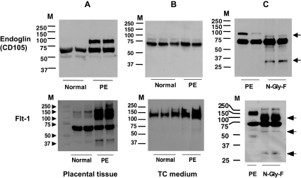
Methods: TCs isolated from normal and PE placentas were cultured under regular (5% CO2/air) and lowered (2% O2/5% CO2/93% N2) oxygen conditions. sEng, sFlt-1, and PlGF productions were determined by ELISA. Protein expressions for Eng, Flt-1, and PlGF in the placental tissues were accessed by immunohistochemical staining and Western blot analysis. Deglycosylated Eng, Flt-1, and PlGF protein expressions in placental tissues were also examined.
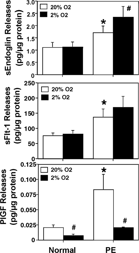
Results: PE TCs produced significantly more sEng, sFlt-1, and PlGF compared with those from normal TCs (P < 0.05). Under lowered oxygen conditions, PE TCs, but not normal TCs, released more sEng and sFlt-1. In contrast, both normal and PE TCs released less PlGF (P < 0.05). Enhanced expressions of Eng and Flt-1, as well as glycosylated Eng and Flt-1, were observed in PE placentas. Immunoblot also revealed that TCs released glycosylated sFlt-1, but not sEng, in culture.
Conclusions: PE TCs produce more sEng, sFlt-1, and PlGF than normal TCs. Lowered oxygen conditions promote sEng and sFlt-1, but reduce PlGF, productions by PE TCs. More glycosylated sEng and sFlt-1 are present in PE placentas. Trophoblasts release glycosylated sFlt-1, but unglycosylated sEng, in culture.
Figures
Similar articles
-
Effects of anti-hypertensive drugs on production of soluble fms-like tyrosine kinase 1 and soluble endoglin from human normal and pre-eclamptic placentas in vitro.Clin Exp Pharmacol Physiol. 2009 Aug;36(8):839-42. doi: 10.1111/j.1440-1681.2009.05155.x. Epub 2009 Feb 10. Clin Exp Pharmacol Physiol. 2009. PMID: 19215236
-
Correlation between soluble endoglin, vascular endothelial growth factor receptor-1, and adipocytokines in preeclampsia.J Clin Endocrinol Metab. 2007 Jul;92(7):2672-9. doi: 10.1210/jc.2006-2349. Epub 2007 Apr 10. J Clin Endocrinol Metab. 2007. PMID: 17426083
-
Heme Oxygenase-1 Is Not Decreased in Preeclamptic Placenta and Does Not Negatively Regulate Placental Soluble fms-Like Tyrosine Kinase-1 or Soluble Endoglin Secretion.Hypertension. 2015 Nov;66(5):1073-81. doi: 10.1161/HYPERTENSIONAHA.115.05847. Epub 2015 Aug 31. Hypertension. 2015. PMID: 26324507
-
[New insights into the pathogenesis of pre-eclampsia: the role of angiogenesis-inhibiting factors].Ned Tijdschr Geneeskd. 2011;155:A2946. Ned Tijdschr Geneeskd. 2011. PMID: 21486506 Review. Dutch.
-
Dysregulation of anti-angiogenic agents (sFlt-1, PLGF, and sEndoglin) in preeclampsia--a step forward but not the definitive answer.J Reprod Immunol. 2009 Nov;82(2):106-11. doi: 10.1016/j.jri.2009.09.001. Epub 2009 Oct 23. J Reprod Immunol. 2009. PMID: 19853925 Review.
Cited by
-
Characterisation of syncytiotrophoblast vesicles in normal pregnancy and pre-eclampsia: expression of Flt-1 and endoglin.PLoS One. 2013;8(2):e56754. doi: 10.1371/journal.pone.0056754. Epub 2013 Feb 20. PLoS One. 2013. PMID: 23437230 Free PMC article.
-
Differential expression of Vegfr-2 and its soluble form in preeclampsia.PLoS One. 2012;7(3):e33475. doi: 10.1371/journal.pone.0033475. Epub 2012 Mar 12. PLoS One. 2012. PMID: 22428059 Free PMC article.
-
Hypertension in Pregnancy: Defining Blood Pressure Goals and the Value of Biomarkers for Preeclampsia.Curr Cardiol Rep. 2016 Dec;18(12):131. doi: 10.1007/s11886-016-0782-1. Curr Cardiol Rep. 2016. PMID: 27837384 Review.
-
Placental expression and methylation of angiogenic factors in assisted reproductive technology pregnancies from India.Epigenomics. 2025 Jan;17(1):21-31. doi: 10.1080/17501911.2024.2438593. Epub 2024 Dec 10. Epigenomics. 2025. PMID: 39655657
-
Glyceryl trinitrate inhibits hypoxia-induced release of soluble fms-like tyrosine kinase-1 and endoglin from placental tissues.Am J Pathol. 2011 Jun;178(6):2888-96. doi: 10.1016/j.ajpath.2011.02.013. Am J Pathol. 2011. PMID: 21641407 Free PMC article.
References
-
- Levine RJ, Maynard SE, Qian C, Lim KH, England LJ, Yu KF, Schisterman EF, Thadhani R, Sachs BP, Epstein FH, Sibai BM, Sukhatme VP, Karumanchi SA 2004 Circulating angiogenic factors and the risk of preeclampsia. N Engl J Med 350:672–683 - PubMed
-
- Levine RJ, Lam C, Qian C, Yu KF, Maynard SE, Sachs BP, Sibai BM, Epstein FH, Romero R, Thadhani R, Karumanchi SA, CPEP Study Group 2006 Soluble endoglin and other circulating antiangiogenic factors in preeclampsia. N Engl J Med [Erratum (2006) 355:1840] 355:992–1005 - PubMed
-
- Maynard SE, Min JY, Merchan J, Lim KH, Li J, Mondal S, Libermann TA, Morgan JP, Sellke FW, Stillman IE, Epstein FH, Sukhatme VP, Karumanchi SA 2003 Excess placental soluble fms-like tyrosine kinase 1 (sFlt1) may contribute to endothelial dysfunction, hypertension, and proteinuria in preeclampsia. J Clin Invest 111:649–658 - PMC - PubMed
-
- Venkatesha S, Toporsian M, Lam C, Hanai J, Mammoto T, Kim YM, Bdolah Y, Lim KH, Yuan HT, Libermann TA, Stillman IE, Roberts D, D’Amore PA, Epstein FH, Sellke FW, Romero R, Sukhatme VP, Letarte M, Karumanchi SA 2006 Soluble endoglin contributes to the pathogenesis of preeclampsia. Nat Med 12:642–649 - PubMed
-
- Park JE, Chen HH, Winer J, Houck KA, Ferrara N 1994 Placenta growth factor. Potentiation of vascular endothelial growth factor bioactivity, in vitro and in vivo, and high affinity binding to Flt-1 but not to Flk-1/KDR. J Biol Chem 269:25646–25654 - PubMed
Publication types
MeSH terms
Substances
Grants and funding
LinkOut - more resources
Full Text Sources
Other Literature Sources
Medical
Miscellaneous
