. 2007 Oct 30;104(44):17329-34.
doi: 10.1073/pnas.0705417104. Epub 2007 Oct 23.

Identifying the subproteome of kinetically stable proteins via diagonal 2D SDS/PAGE

Affiliations

Identifying the subproteome of kinetically stable proteins via diagonal 2D SDS/PAGE

Ke Xia et al. Proc Natl Acad Sci U S A. .

Abstract

Most proteins are in equilibrium with partially and globally unfolded conformations. In contrast, kinetically stable proteins (KSPs) are trapped by an energy barrier in a specific state, unable to transiently sample other conformations. Among many potential roles, it appears that kinetic stability (KS) is a feature used by nature to allow proteins to maintain activity under harsh conditions and to preserve the structure of proteins that are prone to misfolding. The biological and pathological significance of KS remains poorly understood because of the lack of simple experimental methods to identify this property and its infrequent occurrence in proteins. Based on our previous correlation between KS and a protein's resistance to the denaturing detergent SDS, we show here the application of a diagonal 2D (D2D) SDS/PAGE assay to identify KSPs in complex mixtures. We applied this method to the lysate of Escherichia coli and upon proteomics analysis have identified 50 nonredundant proteins that were SDS-resistant (i.e., kinetically stable). Structural and functional analyses of a subset (44) of these proteins with known 3D structure revealed some potential structural and functional biases toward and against KS. This simple D2D SDS/PAGE assay will allow the widespread investigation of KS, including the proteomics-level identification of KSPs in different systems, potentially leading to a better understanding of the biological and pathological significance of this intriguing property of proteins.


Conflict of interest statement

The authors declare no conflict of interest.

Figures

Fig. 1.
Fig. 1.
A simple assay for KS involves comparing the migration of identical unheated (U) and boiled (B) protein samples. Boiled and unheated samples of streptavidin, a KSP, will migrate differently on SDS/PAGE, whereas for β2-microglobulin, which is not kinetically stable, they will have the same migration.
Fig. 2.
Fig. 2.
Diagram of D2D SDS/PAGE method. (a) First-dimension SDS/PAGE separation is performed followed by excision of a gel strip containing the relevant lane. (b) The gel strip is incubated for ≈10 min in a boiling water bath of buffer containing 1% SDS. (c) The gel strip is placed above a larger gel, and a second-dimension SDS/PAGE separation is performed. (d) The gel is stained, revealing a diagonal band resulting from the equal migration of nonKSPs in both dimensions of the gel. Because KSPs migrate less in the first (unheated) dimension, they end up migrating to the left of the gel diagonal.
Fig. 3.
Fig. 3.
Analysis of the cellular lysate of E. coli by D2D SDS/PAGE. The separation in both dimensions was performed as described in Fig. 2 and Materials and Methods. The visible spots to the left of the gel diagonal represent the soluble putative KSPs in E. coli.
Fig. 4.
Fig. 4.
Probing the KS of inorganic pyrophosphatase. (a) The unfolding kinetics of the protein was examined at 4.8–6.7 M GuHCl, and the rate constants were determined by fitting to a single exponential function. (b) The log of the rate constants was plotted against GuHCl and fitted to a linear function. Extrapolation of the plot to 0 M GuHCl yielded a native unfolding rate of 6.44 × 10⁻¹¹·sec−1 (i.e., half-life of 346 years).
Fig. 5.
Fig. 5.
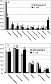
Distribution of protein (a) and enzymatic (b) functions for a nonredundant subset of the E. coli proteome vs. our set of KSPs. (a) The kinetically stable subproteome has significantly more enzymes (P < 0.0001) but fewer transporters (P = 0.0076) and regulators (P = 0.0082). Other changes were not statistically significant at the 95% confidence level. Functional assignments were made by using the E. coli genome and proteome database (16). “Other” refers to the following functions: leader peptides, external origin, cell processes, lipoproteins, pseudogenes, phenotypes, unknown functions, unclassified proteins, and sites. (b) Distribution of the six most common enzyme functions does not show statistically significant differences at the 95% confidence level. Enzyme functions were obtained by using the BRENDA web site (21).
Fig. 6.
Fig. 6.
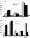
2° (a) and 4° (b) structure distribution of the nonredundant E. coli proteome and its subproteome of KSPs. (a) The KSPs have fewer (P = 0.0034) all α-helical proteins compared with the rest of the E. coli proteome. Structure classifications were made by using the CATH database (17). (b) The KSPs include significantly fewer monomers (P = 0.0002) and significantly more large oligomeric structures with at least five subunits (P < 0.001). Dimers and tetramers occur at approximately the same frequencies in the two sets. 4° structure information was obtained from the PQS Protein Quaternary Structure database (20).

References

    1. Cunningham EL, Jaswal SS, Sohl JL, Agard DA. Proc Natl Acad Sci USA. 1999;96:11008–11014. - PMC - PubMed
    1. Plaza del Pino IM, Ibarra-Molero B, Sanchez-Ruiz JM. Proteins. 2000;40:58–70. - PubMed
    1. Sohl JL, Jaswal SS, Agard DA. Nature. 1998;395:817–819. - PubMed
    1. Kelly JW, Colón W, Lai Z, Lashuel HA, McCulloch J, McCutchen SL, Miroy GJ, Peterson SA. Adv Protein Chem. 1997;50:161–181. - PubMed
    1. Manning M, Colón W. Biochemistry. 2004;43:11248–11254. - PubMed

Publication types

MeSH terms

LinkOut - more resources