. 2008 Feb 1;111(3):1217-26.
doi: 10.1182/blood-2007-07-104133. Epub 2007 Oct 23.

Regulation of angiogenesis through a microRNA (miR-130a) that down-regulates antiangiogenic homeobox genes GAX and HOXA5

Affiliations

Regulation of angiogenesis through a microRNA (miR-130a) that down-regulates antiangiogenic homeobox genes GAX and HOXA5

Yun Chen et al. Blood. .

Abstract

Angiogenesis is critical to tumor progression. The homeobox gene GAX inhibits angiogenesis in vascular endothelial cells (ECs). We have identified a microRNA (miR-130a) that regulates GAX expression and hypothesized that it plays a major role in modulating GAX activity in ECs. A 280-bp fragment from the GAX 3'-untranslated region (3'-UTR) containing 2 miR-130a targeting sites was observed to be required for the rapid down-regulation of GAX expression by serum and proangiogenic factors, whereas the activity of the GAX promoter did not vary with exposure to serum or proangiogenic factors. This same 280-bp sequence in the GAX 3'-UTR cloned into the psiCHECK2-Luciferase vector mediated serum-induced down-regulation of the reporter gene when placed 3' of it. Finally, forced expression of miR-130a inhibits GAX expression through this specific GAX 3'-UTR sequence. A genome-wide search for other possible miR-130a binding sites revealed an miR-130a targeting site in the 3'-UTR of the antiangiogenic homeobox gene HOXA5, the expression and antiangiogenic activity of which are also inhibited by miR-130a. From these data, we conclude that miR-130a is a regulator of the angiogenic phenotype of vascular ECs largely through its ability to modulate the expression of GAX and HOXA5.


Figures

Figure 1
Figure 1
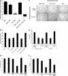
miR-130a, but not the other microRNAs with consensus binding sites in the 3′-UTR of the GAX cDNA, is expressed in HUVECs. (A) GAX promoter activity is not decreased by serum or proangiogenic factors. HUVECs were cotransfected with pGAX-Luciferase and pRL-SV (Renilla luciferase, Dual Luciferase Assay System) as described in “Transfections.” Luciferase activity was normalized to Renilla luciferase activity to control for transfection efficiency. Error bars represent SD. (B,C) Expression of 11 microRNAs in HUVECs in low serum and 2% serum. miR-130a is the only microRNA for which expression is detectable in HUVECs, and its expression is up-regulated by exposure to serum in a dose-dependent fashion. Northern blots were performed as described in “Northern blots” to detect these microRNAs. (D) Location of the 2 miR-130a consensus binding sequences in the GAX 3′-UTR.
Figure 2
Figure 2
The GAX 3′-UTR confers responsiveness to serum and proangiogenic factors in ECs. (A) Construction of the psiCHECK2-GAX-3′-UTR. The 280-bp sequence in the GAX 3′-UTR containing 2 miR-130a consensus sequences was inserted into the vector at the 3′ end of the hRluc (Renilla luciferase) reporter gene. hRluc indicates Renilla reniformis luciferase. (B) Serum and proangiogenic factors suppress luciferase activity in HUVECs transfected with psiCHECK2-GAX-3′-UTR compared with psiCHECK2 empty vector. HUVECs were transduced with either psiCHECK2-GAX-3′-UTR or psiCHECK2 (control) and then incubated in 0.1% FBS, in 0.1% FBS supplemented with 10 ng/mL of either VEGF, bFGF, or TNF-α, or in 2% FBS. Renilla luciferase activities were measured as described in “Dual luciferase reporter assays.” In parallel, identical passaged HUVECs from the same split were transfected with psiCHECK2 empty vector and subjected to the same conditions. Results are expressed as a ratio of the signal from cells transfected with psiCHECK2-GAX-3′-UTR to the signal from cells transfected with psiCHECK2 empty vector. (C) Inhibition of miR-130a reverses the suppression of luciferase activity by serum in a dose-dependent fashion. HUVECs were treated as in B but were cotransfected with increasing concentrations of miR-130a inhibitor. A dose-dependent reversal of the down-regulation of reporter activity attributable to serum was observed. Error bars represent SEM.
Figure 3
Figure 3
The rapid down-regulation of GAX expression after exposure to mitogens requires the miR-130a sequences in the GAX 3′-UTR. (A) GAX mRNA expression is down-regulated by mitogens. Quiescent HUVECs incubated in LSM (0.1% FBS) were exposed to mitogens or conditioned medium from MCF7 cells, incubated overnight, and then harvested for isolation of total mRNA, which was subjected to real-time QRT-PCR. Mitogens strongly down-regulated GAX mRNA expression. (B) Mitogens down-regulate endogenous GAX protein expression. HUVECs were incubated in either 0.1% FBS or 10% FBS overnight and then harvested for total protein, which was subjected to Western blot with anti-Flag antibody. Bands were subjected to densitometry and normalized to α-tubulin levels miR-130a. (C,D) Down-regulation of GAX by mitogens depends on the presence of miR-130a sequences in its 3′-UTR. HUVECs were transfected with either pcDNA3.1-GAX-3′-UTR or pcDNA3.1-GAX, incubated overnight in 0.1%, 2%, or 10% FBS, and then fixed for immunofluorescence (C) or harvested for Western blot (D) with anti-Flag antibodies. In panel C, the photomicrograph was taken at 400× magnification; fluorophores and confocal microscope are described in “Immunofluorescence.” Removing the 3′-UTR sequence almost completely abolishes the down-regulation of GAX attributable to serum stimulation. Error bars represent SEM.
Figure 4
Figure 4
miR-130a down-regulates GAX expression in HUVECs. (A) miR-130a down-regulates GAX mRNA expression. HUVECs were transfected with either empty vector (pcDNA3.1) or miR-130a expression vector (pcDNA3.1-miR-130a) and then incubated overnight in LSM ( = 0.1% FBS), after which total RNA was isolated for QRT-PCR. miR-130a expression markedly down-regulated the expression of endogenous GAX. (B) Representative Western blot of protein from the same experiment as in A. Band intensity was measured by densitometry and normalized to α-tubulin levels. Endogenous GAX protein is also down-regulated by miR-130a. * indicate P < .01. (C,D) Inhibition of GAX expression attributable to miR-130a depends on the presence of the miR-130a binding sites. HUVECs were cotransfected with either pcDNA3.1 or pcDNA3.1-miR-130a plus either pcDNA3.1 or pcDNA3.1-GAX-3′-UTR. Exogenous GAX protein from these plasmids was detected using anti-Flag antibody as described in “miR-130a binding sites in the GAX 3′-UTR mediate serum-induced down-regulation.” Down-regulation of GAX expression by miR-130a was only observed with the construct containing the 2 miR-130a consensus sequences in the 3′-UTR. (C = QRT-PCR; D = Western blot.) (E) Inhibition of miR-130a blocks the down-regulation of GAX attributable to mitogens. HUVECs were transfected with 0 to 100 nM mir-130a inhibitor and incubated in fresh culture medium overnight, after which they were placed in 0.1% FBS or 2% RBS medium for 6 hours. Total RNA was isolated for QRT-PCR as described in insert section. Error bars represent SEM.
Figure 5
Figure 5
miR-130a antagonizes the antiangiogenic activity of GAX. (A) miR-130a antagonizes G0/G1 cell-cycle arrest attributable to GAX depending on the presence of its consensus sequences in the GAX 3′-UTR. HUVECs rendered quiescent by serum starvation overnight were transfected with empty vector or pcDNA-3.1-miR-130a and either pcDNA3.1-GAX or pcDNA3.1-GAX-3′-UTR as for the tube formation experiment and exposed to 10% FBS for 24 hours, after which cells were harvested, stained with PI, and subjected to flow cytometry to determine cell-cycle distribution. By definition, the change in the G0/G1 fraction for the empty vector control is 0, and the G0/G1 fraction for the vector control was 0.38 in the experiment displayed. Statistical significance was determined by one-way ANOVA, and both the miR-130a and GAX-3′-UTR + miR-130a groups showed a statistically significant (P < .01) change from the others. (B) miR-130a antagonizes the antimigration activity of GAX. HUVECs were cotransfected with either pcDNA3.1-GAX-3′-UTR or pcDNA3.1-GAX plus either pcDNA3.1-miR130a or pcDNA3.1 control empty vector and plated 18 hours later onto 8.0 μm pore size polycarbonate membranes in 24-well plates and allowed to attach, after which migration assays were carried out as described in “Migration and tube formation assays.” Cells were counted in 5 hpf per well (*P < .01 compared with empty vector control). (C) miR-130a antagonizes the antiangiogenic activity of GAX. HUVECs were cotransfected with either empty vector or pcDNA3.1-miR-130a and pcDNA3.1-GAX-3′-UTR or pcDNA3.1-GAX and then incubated overnight, after which tube formation assays were carried out. The ratio of cotransfection was 2:1 miR-130a:GAX expression construct. Tube counts were determined as described in “Migration and tube formation assays.” (D) Quantification of tube number per low-powered field (tubes/LPF; *P < .01). (E and F) An inhibitor of miR-130a reverses its antagonism of GAX activity. Experiments described in panels C and D were repeated, but with the addition of 100 nM miR-130a inhibitor. * indicates P < .01. Error bars represent SEM.
Figure 6
Figure 6
miR-130a down-regulates HOXA5 expression. (A) Mitogens down-regulate HOXA5 expression in HUVECs. HUVECs were transfected with full-length HOXA5 (pcDNA3.1-HOXA5), incubated overnight in either 0.1% FBS or 2.0% FBS, and then subjected to immunofluorescence with anti-Flag antibody. The photomicrograph was taken at 400× magnification; fluorophores and confocal microscope are described in “Immunofluorescence.” As with GAX and consistent with previously reported work, serum resulted in the down-regulation of HOXA5 expression.(B) miR-130a down-regulates HOXA5 expression. HUVECs were cotransfected with either pcDNA3.1 or pcDNA3.1-miR-130a plus pcDNA3.1-HOXA5, incubated overnight, and then harvested for Western blot with anti-Flag antibody. miR-130a strongly down-regulates HOXA5 expression. (C) Quantification. Western blots were subjected to densitometry and HOXA5 band densities normalized to those of the corresponding α-tubulin bands. Error bars represnt SEM.
Figure 7
Figure 7
miR-130a also antagonizes the antiangiogenic activity of HOXA5. (A) miR-130a antagonizes the antiangiogenic activity of HOXA5. HUVECs were cotransfected with either empty vector or pcDNA3.1-miR-130a and pcDNA3.1-HOXA5 and then incubated overnight before being subjected to tube formation assays. The ratio of cotransfection was 2:1 miR-130a:HOXA5 expression construct. Tube counts were determined as described in “Migration and tube formation assays.” (B) Quantification of tubes/LPF (*P < .01). Error bars represent SEM.

References

    1. Folkman J. Role of angiogenesis in tumor growth and metastasis. Semin Oncol. 2002;29:15–18. - PubMed
    1. Hanahan D, Folkman J. Patterns and emerging mechanisms of the angiogenic switch during tumorigenesis. Cell. 1996;86:353–364. - PubMed
    1. Bergers G, Benjamin LE. Tumorigenesis and the angiogenic switch. Nat Rev Cancer. 2003;3:401–410. - PubMed
    1. Folkman J. Angiogenesis inhibitors: a new class of drugs. Cancer Biol Ther. 2003;2:S127–S133. - PubMed
    1. Boudreau N, Andrews C, Srebrow A, et al. Induction of the angiogenic phenotype by Hox D3. J Cell Biol. 1997;139:257–264. - PMC - PubMed

Publication types