. 2007 Oct 24;2(10):e1063.
doi: 10.1371/journal.pone.0001063.

Protection of rhesus monkeys by a DNA prime/poxvirus boost malaria vaccine depends on optimal DNA priming and inclusion of blood stage antigens

Affiliations

Protection of rhesus monkeys by a DNA prime/poxvirus boost malaria vaccine depends on optimal DNA priming and inclusion of blood stage antigens

Walter R Weiss et al. PLoS One. .

Abstract

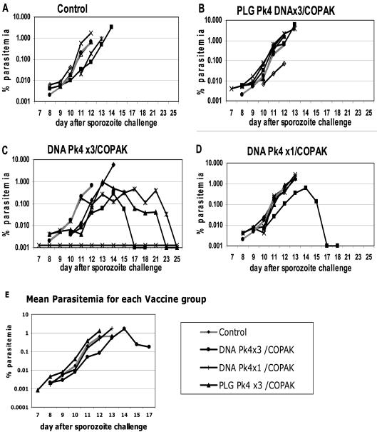
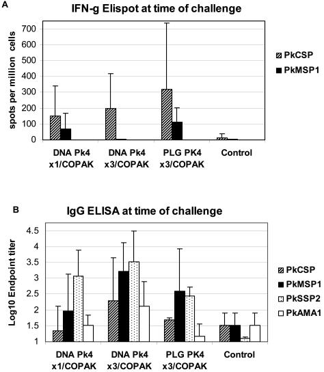
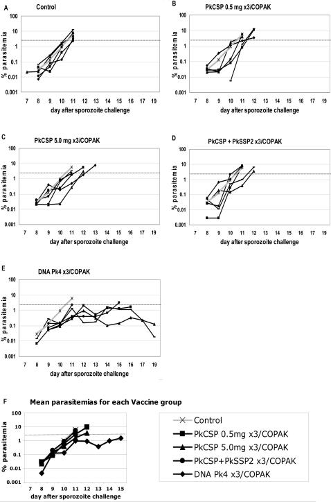
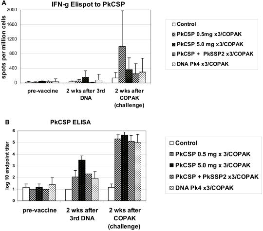
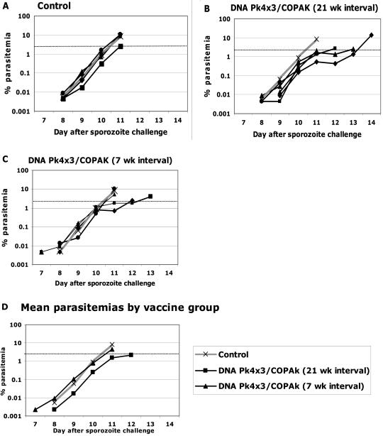
Background: We have previously described a four antigen malaria vaccine consisting of DNA plasmids boosted by recombinant poxviruses which protects a high percentage of rhesus monkeys against Plasmodium knowlesi (Pk) malaria. This is a multi-stage vaccine that includes two pre-erythrocytic antigens, PkCSP and PkSSP2(TRAP), and two erythrocytic antigens, PkAMA-1 and PkMSP-1(42kD). The present study reports three further experiments where we investigate the effects of DNA dose, timing, and formulation. We also compare vaccines utilizing only the pre-erythrocytic antigens with the four antigen vaccine.

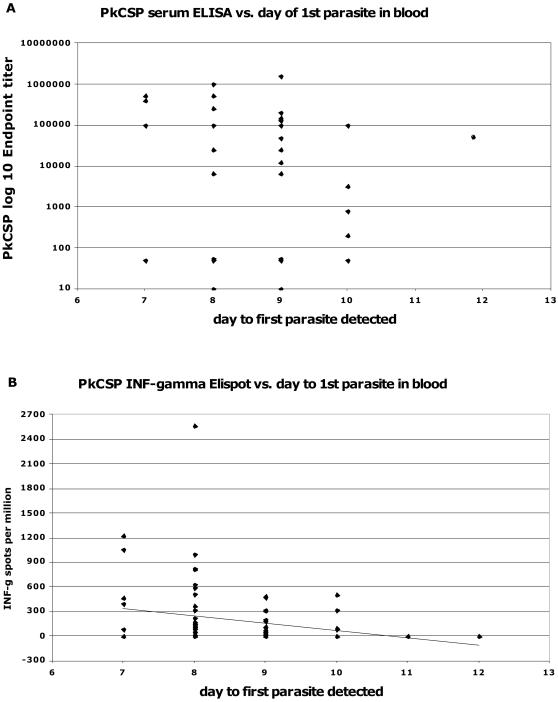
Methodology: In three experiments, rhesus monkeys were immunized with malaria vaccines using DNA plasmid injections followed by boosting with poxvirus vaccine. A variety of parameters were tested, including formulation of DNA on poly-lactic co-glycolide (PLG) particles, varying the number of DNA injections and the amount of DNA, varying the interval between the last DNA injection to the poxvirus boost from 7 to 21 weeks, and using vaccines with from one to four malaria antigens. Monkeys were challenged with Pk sporozoites given i.v. 2 to 4 weeks after the poxvirus injection, and parasitemia was measured by daily Giemsa stained blood films. Immune responses in venous blood samples taken after each vaccine injection were measured by ELIspot production of interferon-gamma, and by ELISA.

Conclusions: 1) the number of DNA injections, the formulation of the DNA plasmids, and the interval between the last DNA injection and the poxvirus injection are critical to vaccine efficacy. However, the total dose used for DNA priming is not as important; 2) the blood stage antigens PkAMA-1 and PkMSP-1 were able to protect against high parasitemias as part of a genetic vaccine where antigen folding is not well defined; 3) immunization with PkSSP2 DNA inhibited immune responses to PkCSP DNA even when vaccinations were given into separate legs; and 4) in a counter-intuitive result, higher interferon-gamma ELIspot responses to the PkCSP antigen correlated with earlier appearance of parasites in the blood, despite the fact that PkCSP vaccines had a protective effect.


Conflict of interest statement

Competing Interests: The authors have declared that no competing interests exist.

Figures

Figure 1
Figure 1. Panels A–D show the % parasitemia for individual monkeys by vaccine group according to the day after sporozoite challenge for Experiment #1.
Data shows the first day parasites were detected and continues until the animal was drug treated when parasitemia exceeded 1% ( 3 animals inadvertently treated at lower parasitemias have open symbols). For comparison, in each panel the grey line shows the mean parasitemia for the Control group. In panel C, one animal never became parasitemic as indicated by x-x. Panel E shows the geometric mean parasitemias for vaccine groups for all days in which at least three animals had not been drug treated. (The monkey from the DNA Pk4×3/COPAK group which did not become infected was excluded from the average).
Figure 2
Figure 2. Panel A. Interferon-γ ELIspot was tested for only two antigens: PkCSP and PkMSP1.
Spots in medium controls were subtracted from antigen test wells, and the results averaged. Priming with DNA on PLG gave a stronger interferon-γ response than DNA in PBS but this was not significant (p = 0.58). Panel B. Geometric mean antibody titers by ELISA at time of sporozoite challenge for each of the four malaria vaccine antigens. Priming with 3 doses of DNA gave higher antibody levels than did a single DNA priming dose for PkCSP, PkMSP1 and PkAMA1 antigens (p<0.05). Priming with 3 doses of DNA in PBS produced higher serum antibody titers than did priming with 3 doses of DNA on PLG for PkCSP, PkSSP2, and PkAMA1 antigens (p<0.05).
Figure 3
Figure 3. Panels A–E show the % parasitemia for individual monkeys by vaccine group according to the day after sporozoite challenge for Experiment #2.
Data shows the first day parasites were detected and continues until the animal was treated with anti-malarial drugs. For comparison, in each panel the grey line shows the mean parasitemia for the Control group. Panel F shows the geometric mean parasitemias for all the vaccine groups for all days in which at least three animals had not been drug treated.
Figure 4
Figure 4. Panel A shows interferon-γ ELIspot response to PkCSP by vaccine group prior to vaccination, after the third DNA vaccination, and after the COPAK boost.
After the third DNA vaccination, the PkCSP 5.0 mg group has the highest response but this is not significantly greater than the PkCSP 0.5 mg group (p = 0.11). After the third DNA vaccination monkeys receiving both PkCSP and PkSSP2 immunizations had significantly lower responses to PkCSP than did animals receiving only PkCSP DNA (p = 0.04). After the COPAK boost, monkeys primed with low dose PkCSP 0.5 mg DNA had the highest response interferon-γ response (p = 0.08 vs high dose PkCSP, p = 0.04 vs Pk4 vaccine, p = 0.04 vs PkCSP+PkSSP2). Panel B shows IgG responses at the three time points. After three DNA vaccinations, serum titers were highest in the high dose DNA PkCSP 5.0 mg group (p = 0.01). After boosting with recombinant COPAK virus, monkeys primed with the high dose DNA had the highest serum IgG titers but this was not significantly different from the other vaccine groups.
Figure 5
Figure 5. Panels A–C show the % parasitemias for individual monkeys by vaccine group according to the day after sporozoite challenge for Experiment #3.
Data shows the first day parasites were detected and continues until the animal was treated with anti-malaria drugs. For comparison, in each panel the grey line shows the mean parasitemia for the Control group. Panel D shows the geometric mean parasitemias for all the vaccine groups for all days in which at least three animals had not been drug treated. For each day 8–11 the mean parasitemia for the group receiving the booster dose at the 21 week interval was lower than the other groups (p<0.05, Student's T test).
Figure 6
Figure 6. Immune response to PkCSP antigen at time of challenge for individual animals by day to first parasite seen in blood.
Data from Experiments #1, 2, and 3 are plotted. Data from Control animals are not included. Panel A shows PkCSP endpoint ELISA titers where there is no significant correlation. Panel B shows PkCSP interferon-γ ELIspot titers with a linear regression line included. There is a significant negative correlation (p = 0.04), animals with lower ELIspot responses having longer times to the appearance of first parasite in the blood.

References

    1. Ballou WR. Malaria vaccines in development. Expert Opin Emerg Drugs. 2005;10:489–503. - PubMed
    1. Ballou WR, Arevalo-Herrera M, Carucci D, Richie TL, Corradin G, et al. Update on the clinical development of candidate malaria vaccines. Am J Trop Med Hyg. 2004;71:239–247. - PubMed
    1. Enosse S, Dobano C, Quelhas D, Aponte JJ, Lievens M, et al. RTS,S/AS02A Malaria Vaccine Does Not Induce Parasite CSP T Cell Epitope Selection and Reduces Multiplicity of Infection. PLoS Clin Trials. 2006;1:e5. - PMC - PubMed
    1. Alonso PL, Sacarlal J, Aponte JJ, Leach A, Macete E, et al. Efficacy of the RTS,S/AS02A vaccine against Plasmodium falciparum infection and disease in young African children: randomised controlled trial. Lancet. 2004;364:1411–1420. - PubMed
    1. Alonso PL, Sacarlal J, Aponte JJ, Leach A, Macete E, et al. Duration of protection with RTS,S/AS02A malaria vaccine in prevention of Plasmodium falciparum disease in Mozambican children: single-blind extended follow-up of a randomised controlled trial. Lancet. 2005;366:2012–2018. - PubMed

Publication types