. 2007 Oct 24;2(10):e1082.
doi: 10.1371/journal.pone.0001082.

Conformational reorganization of the SARS coronavirus spike following receptor binding: implications for membrane fusion

Affiliations

Conformational reorganization of the SARS coronavirus spike following receptor binding: implications for membrane fusion

Daniel R Beniac et al. PLoS One. .

Abstract

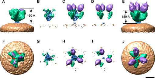
The SARS coronavirus (SARS-CoV) spike is the largest known viral spike molecule, and shares a similar function with all class 1 viral fusion proteins. Previous structural studies of membrane fusion proteins have largely used crystallography of static molecular fragments, in isolation of their transmembrane domains. In this study we have produced purified, irradiated SARS-CoV virions that retain their morphology, and are fusogenic in cell culture. We used cryo-electron microscopy and image processing to investigate conformational changes that occur in the entire spike of intact virions when they bind to the viral receptor, angiotensin-converting enzyme 2 (ACE2). We have shown that ACE2 binding results in structural changes that appear to be the initial step in viral membrane fusion, and precisely localized the receptor-binding and fusion core domains within the entire spike. Furthermore, our results show that receptor binding and subsequent membrane fusion are distinct steps, and that each spike can bind up to three ACE2 molecules. The SARS-CoV spike provides an ideal model system to study receptor binding and membrane fusion in the native state, employing cryo-electron microscopy and single-particle image analysis.


Conflict of interest statement

Competing Interests: The authors have declared that no competing interests exist.

Figures

Figure 1
Figure 1. Imaging SARS-CoV, Vero E6 cells, and ACE2.
(A) Vero E6 cells stained with propidium iodide. Light microscopy shows a syncytium (white arrow) in the center of the field of view (top), viewed under propidium iodide fluorescence (middle), and an overlay of both images (bottom). (B) Immuno-EM with 10 nm gold confirmed the attachment of ACE2 to the spikes. (C) Cryo-EM field of view of SARS-CoV decorated with ACE2, and select two-dimensional class averages of the SARS spike-ACE2 complex (D). Cryo-EM reconstruction of the SARS spike (E), and SARS spike-ACE2 complex (F), viewed from side and end-on perspectives (left, right). Color scheme: ACE2, violet; spike, green; stalk, blue; envelope, beige; nucleocapsid, red. Scale bars: (A) 50 µm, (B,C) 1000 Å, (D–F) 100 Å.
Figure 2
Figure 2. Estimate of resolution of the SARS spike-ACE2 complex.
The resolution was estimated by Fourier shell correlation (FSC) between two half reconstructions of the SARS spike-ACE2 complex. The resolution was estimated to be 18.5 Å using the 0.5 FSC criteria (red dotted red line).
Figure 3
Figure 3. Cryo-EM difference maps.
The cryo-EM reconstruction of the SARS spike (A,F) was subtracted from the SARS spike-ACE2 complex (E,J). The positive component attributed to the SARS spike in (B,G) indicates a re-arrangement in the S2 core, and the positive component attributed the SARS-CoV–ACE2 complex shows the addition of ACE2 and an exterior re-arrangement in S1 (D,I). The net difference map is presented in (C,H). The structures are presented from a side perspective (A–E), and end-on perspective (F–J). The arrows in (F) and (J) illustrate the mass reorganization that occurs in the central axis of the spike where one small central blob splits into three nubs. In (A,F) the region on the spike adjacent to ACE2 which corresponds to the receptor binding domain, has been highlighted with a dotted line and is colored purple. The color scheme used for the cryo-EM structure is the same as in Figure 1. Scale bar, 100 Å.
Figure 4
Figure 4. Merging cryo-EM and atomic resolution 3D structures.
The atomic resolution structures; PDB ID code, 2AJF (ACE2, blue; receptor-binding domain, red) and PDB ID code, 2FXP (yellow) were docked within the SARS spike-ACE2 complex using SITUS. The SARS spike-ACE2 complex is shown from (A) side and (B) end-on perspectives, the arrow points to the C-terminus of ACE2. The color scheme used for the cryo-EM structure is the same as in Figure 1. Scale bar, 100 Å.

Similar articles

Cited by

References

    1. Dutch RE, Jardetzky TS, Lamb RA. Virus membrane fusion proteins: biological machines that undergo a metamorphosis. Biosci Rep. 2000;20:597–612. - PubMed
    1. Hakansson-McReynolds S, Jiang S, Rong L, Caffrey M. Solution structure of the severe acute respiratory syndrome-coronavirus heptad repeat 2 domain in the prefusion state. J Biol Chem. 2006;281:11965–11971. - PMC - PubMed
    1. Xu Y, Lou Z, Liu Y, Pang H, Tien P, et al. Crystal structure of severe acute respiratory syndrome coronavirus spike protein fusion core. J Biol Chem. 2004;279:49414–49419. - PMC - PubMed
    1. Skehel JJ, Wiley DC. Receptor binding and membrane fusion in virus entry: the influenza hemagglutinin. Annu Rev Biochem. 2000;69:531–569. - PubMed
    1. Kobe B, Center RJ, Kemp BE, Poumbourios P. Crystal structure of human T cell leukemia virus type 1 gp21 ectodomain crystallized as a maltose-binding protein chimera reveals structural evolution of retroviral transmembrane proteins. Proc Natl Acad Sci U S A. 1999;96:4319–4324. - PMC - PubMed

Publication types

Substances