

Two anomalous localizations of mucocele: clinical presentation and retrospective review
- PMID: 17957852
- PMCID: PMC2640024
Two anomalous localizations of mucocele: clinical presentation and retrospective review
Abstract
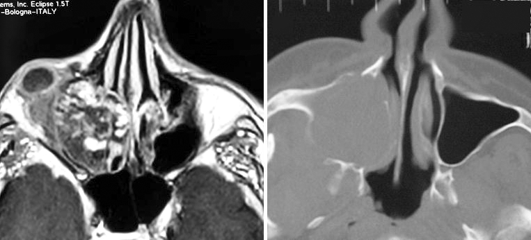
Two Caucasian males (57 and 70 years old) were referred to our attention with parasinus mucoceles, maxillary and frontal mucocele, respectively, that had eroded the orbital rim and caused swelling of the eyelids and brow. Invasion of the orbital space caused several ophthalmic symptoms such as diplopia, proptosis, ptosis, and the formation of a palpable mass. Ophthalmic involvement was the first sign of the mucocele. The mucoceles were completely excised through a skin incision and the diseased mucosa of the sinuses was removed: endonasal fibre optic surgery and the Caldwell-Luc procedure were used in the patient with maxillary mucocele. The cases are described with retrospective review.
Due uomini caucasici (di 57 e 70 anni) sono giunti alla nostra osservazione per mucocele dei seni paranasali, rispettivamente mucocele mascellare e frontale, con erosione del bordo orbitale e tumefazione palpebrale e del sopracciglio. L’invasione della cavità orbitale aveva causato molti disturbi oftalmici quali diplopia, proptosi, ptosi e la formazione di una massa palpabile. Il coinvolgimento oftalmico è stato il primo segno del mucocele. I mucoceli sono stati completamente asportati mediante un’incisione cutanea e la mucosa patologica dei seni è stata rimossa chirurgicamente: nel paziente affetto da mucocele mascellare le procedure chirurgiche sono state una chirurgia per via endoscopica mediante l’uso di fibre ottiche e tecnica tipo Caldwell-Luc. Vengono descritti i casi con una revisione della letteratura.
Figures


Similar articles
-
Clinical manifestations and management of orbital mucoceles: the role of ophthalmologists.Jpn J Ophthalmol. 2005 May-Jun;49(3):239-45. doi: 10.1007/s10384-004-0174-8. Jpn J Ophthalmol. 2005. PMID: 15944832
-
[A case of maxillary sinus mucocele with orbital involvement].Kulak Burun Bogaz Ihtis Derg. 2007;17(5):290-3. Kulak Burun Bogaz Ihtis Derg. 2007. PMID: 18187990 Turkish.
-
Orbital complications of infected mucocele in the paranasal sinuses.Auris Nasus Larynx. 2020 Dec;47(6):990-995. doi: 10.1016/j.anl.2020.05.012. Epub 2020 Jun 11. Auris Nasus Larynx. 2020. PMID: 32536502
-
Frontoethmoidal mucocele presenting with ocular manifestations.Clin Exp Optom. 2020 Sep;103(5):610-617. doi: 10.1111/cxo.13006. Epub 2019 Nov 26. Clin Exp Optom. 2020. PMID: 31773805 Review.
-
Mucoceles of the sphenoidal sinus: a report of four cases and review of the literature.B-ENT. 2005;1(4):181-5. B-ENT. 2005. PMID: 16429750 Review.
Cited by
-
Orbital myeloid sarcoma (chloroma): Report of 2 cases and literature review.Am J Ophthalmol Case Rep. 2020 Jul 11;19:100806. doi: 10.1016/j.ajoc.2020.100806. eCollection 2020 Sep. Am J Ophthalmol Case Rep. 2020. PMID: 32775766 Free PMC article.
References
-
- Shields JA, Shields CL. Orbital cysts of childhood – classification, clinical features, and management. Surv Ophthal 2004;49:281-99. - PubMed
-
- Rootman J, Chang W, Jones D. Distribution and differential diagnosis of orbital disease. In: Rootman J, editor. Disease of the orbit. A multidisciplinary approach. Philadelphia: Lippincott, Williams and Wilkins; 2003. p. 53-84.
-
- Schick U, Hassler W. Neurosurgical management of orbital inflammations and infections. Acta Neurochir 2004;146:571-80. - PubMed
-
- Khong JJ, Malhotra R, Wormald PJ, Selva D. Endoscopic sinus surgery for paranasal sinus mucocoele with orbital involvement. Eye 2004;18:877-81. - PubMed
-
- Wang TJ, Liao SL, Jou JR, Lin LK. Clinical manifestation of orbital mucocele: the role of Ophthalmologists. Jpn J Ophthalmol 2005;49:239-45. - PubMed
Publication types
MeSH terms
LinkOut - more resources
Full Text Sources