. 2007 Oct 24:2:20.
doi: 10.1186/1749-8104-2-20.

Metamorphosis of an identified serotonergic neuron in the Drosophila olfactory system

Affiliations

Metamorphosis of an identified serotonergic neuron in the Drosophila olfactory system

Bidisha Roy et al. Neural Dev. .

Abstract

Background: Odors are detected by sensory neurons that carry information to the olfactory lobe where they connect to projection neurons and local interneurons in glomeruli: anatomically well-characterized structures that collect, integrate and relay information to higher centers. Recent studies have revealed that the sensitivity of such networks can be modulated by wide-field feedback neurons. The connectivity and function of such feedback neurons are themselves subject to alteration by external cues, such as hormones, stress, or experience. Very little is known about how this class of central neurons changes its anatomical properties to perform functions in altered developmental contexts. A mechanistic understanding of how central neurons change their anatomy to meet new functional requirements will benefit greatly from the establishment of a model preparation where cellular and molecular changes can be examined in an identified central neuron.

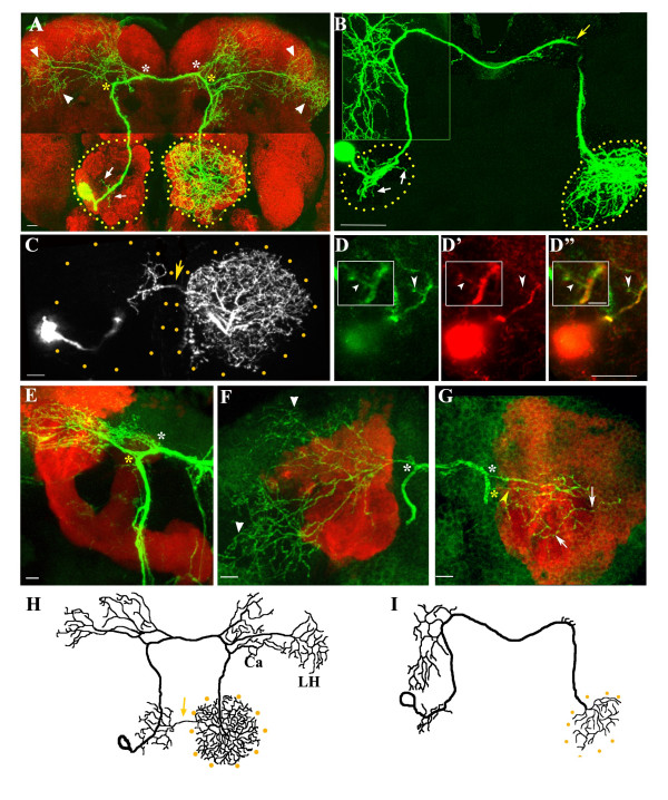
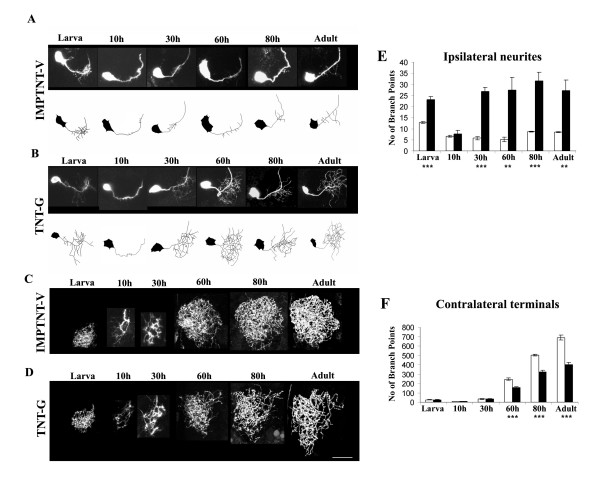
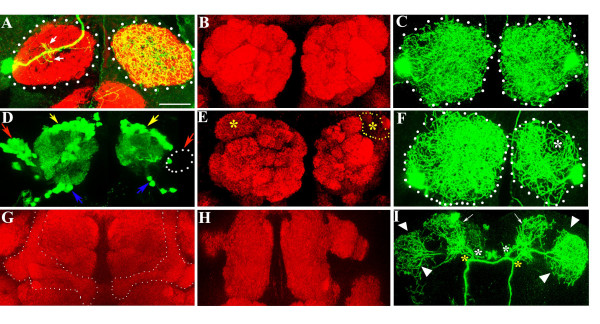
Results: In this study, we examine a wide-field serotonergic neuron in the Drosophila olfactory pathway and map the dramatic changes that it undergoes from larva to adult. We show that expression of a dominant-negative form of the ecdysterone receptor prevents remodeling. We further use different transgenic constructs to silence neuronal activity and report defects in the morphology of the adult-specific dendritic trees. The branching of the presynaptic axonal arbors is regulated by mechanisms that affect axon growth and retrograde transport. The neuron develops its normal morphology in the absence of sensory input to the antennal lobe, or of the mushroom bodies. However, ablation of its presumptive postsynaptic partners, the projection neurons and/or local interneurons, affects the growth and branching of terminal arbors.

Conclusion: Our studies establish a cellular system for studying remodeling of a central neuromodulatory feedback neuron and also identify key elements in this process. Understanding the morphogenesis of such neurons, which have been shown in other systems to modulate the sensitivity and directionality of response to odors, links anatomy to the development of olfactory behavior.


Figures

Figure 1
Figure 1
Architecture of the CSD neuron. The neuron is marked by a flipout in the RN2-Flp, Tub-FRT-CD2-FRTGal4, UAS-mCD8GFP strain, resulting in GFP expression in a single CSD neuron. (a) Only one of the pair of CSD neurons is labeled with antibodies to GFP (green) and the brain neuropile is stained with mAbnc82 (red). 1 μm sections at appropriate levels are stacked to visualize the cell body and ipsilateral dendrites (arrows), the branches to the higher centers (asterisks), projections to the lateral protocerebrum (arrowheads) in both hemispheres, and the presynaptic terminals in the contralateral antennal lobe (demarcated by yellow dots). (b) A single CSDn visualized in the brain of the third instar larva. Dendritic arbors ramify in the ipsilateral lobe (white arrows), branch extensively at the ipsilateral higher centers (inset) and send only a few arbors on the contralateral side (yellow arrow). The terminal arbors are extensive within the contralateral antennal lobe (demarcated with dotted lines). (c) In the adult, the terminal arbors of the CSDn arborize within the contralateral lobe and also send a branch in the antennal commissure (yellow arrow) to the ipsilateral antennal lobe. (d-d") Nod::lacZ localization in the ipsilateral dendrites (arrowheads) is visualized by anti-β-galactosidase staining (d', d", red). Inset in (d-d") shows Nod::lacZ localization in an ipsilateral neurite (arrowhead). (e-g) RN2-Flp, Tub-FRT-CD2-FRTGal4, UAS-mCD8GFP/mb247-DsRed animal stained with antibodies against GFP and visualized in the green (GFP) and red (ds-red) channels of the confocal microscope. Branches exit the main trunk in both hemispheres (yellow and white asterisks) and traverse anterior to the mushroom bodies to terminate within the higher centers (arrowheads in (f)). (g) A few sections are stacked to show the arborization to the calyx from the contralateral branches (arrows); the arrowhead indicates the branchpoint to the calyx neurites. (h, i) Schematic diagrams of the CSD neuron showing its projection patterns in the adult (h) and the larvae (i). The arrow in (h) indicates the crossover of presynaptic terminals to the lobe ipsilateral to the soma. Ca, Calyx; LH, lateral horn. Scale bar in (d) inset = 5 μm; for all other figures = 10 μm.
Figure 2
Figure 2
Serotonin immunoreactivity and the effect of serotonin depletion on the CSDn. (a-d) Staining using anti-serotonin antibodies (red) in different regions of the CSDn (green); (a) cell body, (b) contralateral terminals, (c) branches in the ipsilateral and (d) contralateral higher centers (arrows highlight puncta of serotonin immunoreactivity). (e-h) ddcts2/ddcts2; RN2-Flp, Tub-FRT-CD2-FRTGal4, UAS-mCD8GFP/+ animals grown at restrictive temperature show a strong reduction in serotonin immunoreactivity within the antennal lobe (e). The presynaptic terminals from a single CSDn (f), as well as the ipsilateral (g) and contralateral (h) arbors over the higher centers (arrowheads) do not show any appreciable change in branching pattern. Scale bar in all figures = 10 μm
Figure 3
Figure 3
Re-modeling of the CSD neuron during pupal life. (a-d) Developmental changes in different regions of the CSD neuron; (a) cell body and dendrites within the ipsilateral lobe, (b) presynaptic terminals in the contralateral lobe, (c) branches in the ipsilateral higher centers, and (d) branches in the contralateral higher centers. The regions are shown at (i) 0 hours APF, (ii) 10 hours APF, (iii) 40 hours APF, (iv) 80 hours APF and (v) adult. Scale bar = 5 μm in (i) and (ii); 10 μm in (iii); 40 μm in (iv) and (v). Yellow arrows indicate branches being pruned while green arrows point to the new sprouts. The branching has been quantified as described in Additional file 3 and plotted in histograms in (a(vi)), (b(vi)), (c(vi)) and (d(vi)). The number of branch points is represented at different time points; bars indicate the mean and standard error of mean of five preparations in each case. (e) Infiltration of glial cells at the sites of pruning of the CSD neuron at (i) 10 hours and (ii) 20 hours APF. The glial cells are visualized by staining with an antibody against Repo (red). The means and standard error of mean of the number of cells in five preparations are represented in the histogram in (e(iv)).
Figure 4
Figure 4
Role of ecdysterone signaling in remodeling of the CSD neuron. (a, c, d) CSDn profiles visualized in RN2-Flp, Tub-FRT-CD2-FRT-Gal4, UAS-mCD8GFP/UAS-EcRW650A animals at different developmental stages. (b) RN2-Flp, Tub-FRT-CD2-FRT-Gal4, UAS-mCD8GFP/+ control at 20 hours APF; scale bar for all images = 30 μm. (a) In the larvae, branching within the antennal lobe (red arrow), the ipsilateral higher centers (arrowhead) and the terminals are comparable to that of the wild type (Figure 1b). Branches at the contralateral higher centers (yellow arrows) appear more elaborate than that of normal controls (Figure 1b). Staining with antibodies against the EcR-B1 isoform (red in inset) and anti-GFP (green in inset) demonstrated that the CSDn soma (arrows in inset) expressed EcR-B1. At 20 hours APF, pruning has occurred in the normal CSDn (b) and branching within the antennal lobes (demarcated with dotted lines), the ipsilateral (arrowheads) and contralateral (arrows) are greatly reduced. CSDn expressing a DN-EcR transgene (c) does not undergo pruning and the neuronal architecture resembles that of the larva. In the adult (d), the pattern continues to be 'larva-like'. However, the footprint of the terminal arbors occupies a larger area than in the larva (demarcated with yellow dots) and many of the arbors appear blebby (arrowheads in inset in (d)).
Figure 5
Figure 5
Targeted expression of TeTxLC, Shits1, Kir2.1 and Glued-DN affects the architecture of the CSDn. Adult brains were stained with anti-GFP (green) and mAbnc82 (red). (a, g) Wild-type (WT) CSDn showing the cell body with dendrites in the ipsilateral lobe (arrows in (a)) and terminal arbors in the contralateral lobe (g). (b, h) The neuron expressing inactive tetanus toxin (UAS-IMPTNT-V/+; RN2-Flp, Tub-FRT-CD2-FRT-Gal4, UAS-CD8GFP/+) have dendrites (arrow in (b)) and terminal arbors (h) comparable to WT. (c, i) Ectopic expression of active form of tetanus toxin (UAS-TNT-G/+; RN2-Flp, Tub-FRT-CD2-FRT-Gal4, UAS-CD8GFP/+) results in dramatic increase in ipsilateral dendrites (arrows in (c)) and a marked decrease in contralateral terminals (i). (d, j) UAS-shits1/+; RN2-Flp, Tub-FRT-CD2-FRT-Gal4, UAS-CD8GFP/UAS-shits1 showing greater profusion of dendrites in the ipsilateral lobe (arrows in (d)) and reduced terminal arbors (j). (e, k) RN2-Flp, TubFRT-CD2-FRT-Gal4, UAS-CD8GFP/UAS-EGFPKir2.1 showing greatly increased dendritic branches within the ipsilateral lobe (arrows in (e)) and modest decrease in contralateral terminals (k). (f, l) UAS-Glued-DN/+; RN2-Flp, Tub-FRT-CD2-FRT-Gal4, UAS-CD8GFP/+ showing increased ipsilateral dendrites (arrows in (f)) and a reduction in contralateral terminals (l). Scale bar = 30 μm. (m, n) Number of branch points from the ipsilateral (m) (N = 7) and contralateral (n) (N = 5) arbors were quantified from all the genotypes. Bars represent the mean and standard error of mean of number of branch points. P-values were calculated using the unpaired Student's t-test *P < 0.05, **P < 0.01, ***P < 0.0001.
Figure 6
Figure 6
Effect of expression of TeTxLC on the remodeling of the CSD neuron. The developmental profile of the CSDn in animals where the (a, c) inactivated (UAS-IMPTNT-V/+; RN2-Flp, Tub-FRT-CD2-FRT-Gal4, UAS-CD8GFP/+) and (b, d) activated (UAS-TNT-G/+; RN2-Flp, Tub-FRT-CD2-FRT-Gal4, UAS-CD8GFP/+) forms of TeTxLC have been expressed. Animals where a unilateral flipout had occurred were analyzed in detail at the larva, 10 hour APF, 30 hour APF, 60 hour APF, 80 hour APF and adult stages. Scale bar for all images = 30 μm. The dendritic trees from preparations where inactivated (a) and activated (b) TeTxLC have been ectopically expressed are schematized. The presynaptic terminals in the contralateral lobe where IMPTNT-V and TNT-G were expressed are shown in (c) and (d), respectively. The branch points were quantified as described in Additional file 3 and represented in the histograms in (e) for the ipsilateral lobe and (f) for the contralateral lobe. Five preparations were analyzed at each data point; white bars represent UAS-IMPTNT-V/+; RN2-Flp, Tub-FRT-CD2-FRT-Gal4, UAS-CD8GFP/+; black bars represent UAS-TNTG/+; RN2-Flp, TubFRT-CD2-FRTGal4, UAS-CD8GFP/+. P-values were calculated using the unpaired Student's t-test; **P < 0.001, ***P < 0.0001.
Figure 7
Figure 7
Effect of ablation of subsets of neurons within the olfactory circuit on the development of the CSD neuron. (a) Antennal lobes (demarcated with dotted lines) of lz3; +/+; Antp/RN2-Flp, Tub-FRT-CD2-FRT-Gal4, UAS-CD8GFP stained with anti-GFP (green) and mAbnc82 (red). lz3; +/+; Antp/+ animals lack ORNs projecting from the third antennal segment. As in the wild-type (Figure 1a), the CSDn in this genetic background sends a few dendrites into the ipsilateral lobe (small arrows) and terminates by extensive branching in the lobe contralateral to the cell body. The glomeruli are less well demarcated by mAbnc82 staining compared to control brains shown in (b). (c) RN2-Flp, Tub-FRT-CD2-FRT-Gal4, UAS-CD8GFP where terminals of both CSDn are labeled within the antennal lobes. (d-f, h, i) Brains of animals that had been fed with HU, stained with anti-GFP (green) and mAbnc82 (red). (d, e) GH146-Gal4, UAS-GFP. (d) Cell bodies of the PNs lie in three clusters located anterodorsal (blue arrows), posterior (yellow arrows) and lateral (red arrows) to the antennal lobe. A subset of the lateral cluster neurons (dotted region) is absent in one lobe. (e) This results in the absence of a glomerulus denoted with (*-DA1) on the affected side (yellow dotted lines). (f) RN2-Flp, Tub-FRT-CD2-FRT-Gal4, UAS-mCD8GFP animals treated as the GH146-Gal4, UAS-GFP animals described above. Animals with one lobe reduced in size were selected and the terminal arbors from bilateral flip-outs examined. The affected side shows significantly less branching, particularly over the region where the DA1 is positioned (asterisk; compare with (c)). (g) Mushroom bodies are indicated by dotted lines in control (no HU) preparations. (h, i) In HU treated animals where the mushroom bodies were absent (h), the architecture of the CSDn was normal (i). Secondary branches (yellow and white asterisk) that arborize over the calyx of the mushroom bodies (arrows) and the lateral horn (arrowheads) appear normal. Arrows indicate branching at the calyx region and arrowheads the branching at the lateral horn. Scale bar = 20 μm.

Similar articles

Cited by

References

    1. Komiyama T, Luo L. Development of wiring specificity in the olfactory system. Curr Opin Neurobiol. 2006;16:67–73. doi: 10.1016/j.conb.2005.12.002. - DOI - PubMed
    1. Gao Q, Yuan B, Chess A. Convergent projections of Drosophila olfactory neurons to specific glomeruli in the antennal lobe. Nat Neurosci. 2000;3:780–785. doi: 10.1038/75753. - DOI - PubMed
    1. Vosshall LB, Wong AM, Axel R. An olfactory sensory map in the fly brain. Cell. 2000;102:147–159. doi: 10.1016/S0092-8674(00)00021-0. - DOI - PubMed
    1. Stocker RF. The organization of the chemosensory system in Drosophila melanogaster: a review. Cell Tissue Res. 1994;275:3–26. doi: 10.1007/BF00305372. - DOI - PubMed
    1. Shang Y, Claridge-Chang A, Sjulson L, Pypaert M, Miesenbock G. Excitatory local circuits and their implications for olfactory processing in the fly antennal lobe. Cell. 2007;128:601–612. doi: 10.1016/j.cell.2006.12.034. - DOI - PMC - PubMed

Publication types

MeSH terms

LinkOut - more resources