. 2008 Jan;82(2):817-27.
doi: 10.1128/JVI.01847-07. Epub 2007 Oct 24.

Human astrovirus coat protein inhibits serum complement activation via C1, the first component of the classical pathway

Affiliations

Human astrovirus coat protein inhibits serum complement activation via C1, the first component of the classical pathway

Rheba S Bonaparte et al. J Virol. 2008 Jan.

Abstract

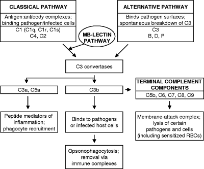
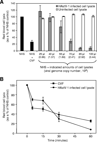
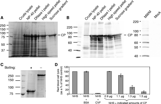
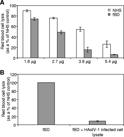
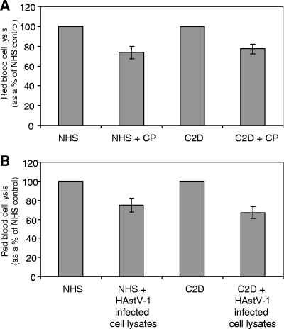
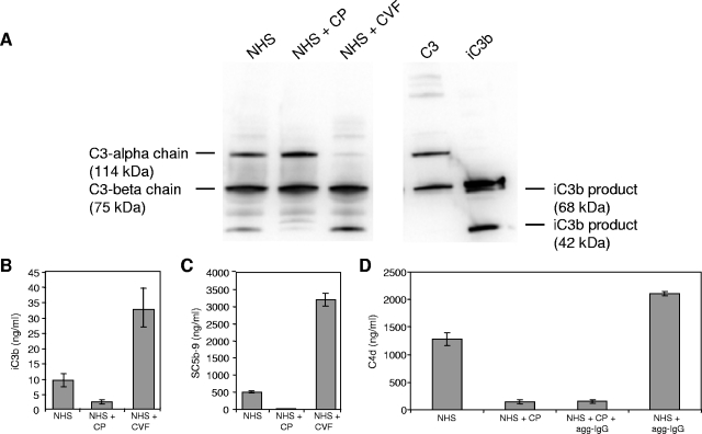
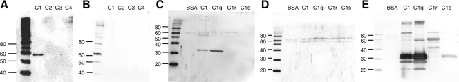
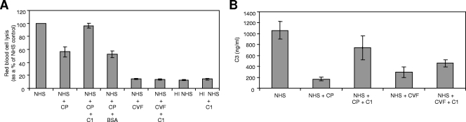
Human astroviruses (HAstVs) belong to a family of nonenveloped, icosahedral RNA viruses that cause noninflammatory gastroenteritis, predominantly in infants. Eight HAstV serotypes have been identified, with a worldwide distribution. While the HAstVs represent a significant public health concern, very little is known about the pathogenesis of and host immune response to these viruses. Here we demonstrate that HAstV type 1 (HAstV-1) virions, specifically the viral coat protein (CP), suppress the complement system, a fundamental component of the innate immune response in vertebrates. HAstV-1 virions and purified CP both suppress hemolytic complement activity. Hemolytic assays utilizing sera depleted of individual complement factors as well as adding back purified factors demonstrated that HAstV CP suppresses classical pathway activation at the first component, C1. HAstV-1 CP bound the A chain of C1q and inhibited serum complement activation, resulting in decreased C4b, iC3b, and terminal C5b-9 formation. Inhibition of complement activation was also demonstrated for HAstV serotypes 2 to 4, suggesting that this phenomenon is a general feature of these human pathogens. Since complement is a major contributor to the initiation and amplification of inflammation, the observed CP-mediated inhibition of complement activity may contribute to the lack of inflammation associated with astrovirus-induced gastroenteritis. Although diverse mechanisms of inhibition of complement activation have been described for many enveloped animal viruses, this is the first report of a nonenveloped icosahedral virus CP inhibiting classical pathway activation at C1.


Figures

FIG. 1.
FIG. 1.
Overview of the pathways of serum complement activation. The main protein factors and their effector actions are indicated.
FIG. 2.
FIG. 2.
HAstV-1 virions suppress complement activity in a hemolytic complement assay, with similar kinetics to those of CVF. (A) Antibody-sensitized sheep RBCs were incubated with 20 μl NHS alone or in the presence of 1 μg CVF (black bars), along with increasing amounts of infected CaCo-2 cell lysates containing HAstV-1 virions (shaded bars) or uninfected CaCo-2 cell lysates (white bars). Hemolysis was standardized to 100% for NHS alone. One microliter of infected cell culture lysate corresponds to 3.44 × 10⁶ genome copies, as determined by real-time RT-PCR. (B) CaCo-2 cell lysates containing HAstV-1 virions (85 μl of cell lysate, corresponding to 2.92 × 10⁸ genome copies) and 1 μg CVF were incubated for 0 to 60 min in the presence of 20 μl NHS. At 0, 5, 15, 30, and 60 min, aliquots were removed and incubated with sensitized sheep RBCs to test hemolytic complement activity. Data are the means from five independent experiments. Error bars denote standard errors of the means (SEM).
FIG. 3.
FIG. 3.
SDS-PAGE and immunoblot analysis of fractions obtained during the sucrose gradient ultracentrifugation step of HAstV-1 CP purification. The first 18 fractions of the gradient were run in SDS-PAGE gels and stained with Coomassie blue (A) or subjected to immunoblotting with antibody to HAstV-1 virions (B). Numbers refer to gradient fractions, with arrows representing the direction of sedimentation. Molecular mass markers (in kDa) are indicated in the middle, while the position of CP is indicated to the right.
FIG. 4.
FIG. 4.
Analysis of HAstV-1 CP purification, oligomerization state, and activity in the hemolytic complement assay. Coomassie blue staining (A) and immunoblot analysis (B) of the CP-containing fraction were performed at each stage of the purification procedure. An IPLB-Sf21 mock-infected cellular lysate was also analyzed by immunoblotting to demonstrate that there was no cross-reactivity to the HAstV-1 antibody. Molecular size markers (MWM; in kDa) are indicated to the left, with the position of CP denoted on the right. (C) Aliquots of sucrose-purified CP were either boiled or not boiled in the presence of 2-mercaptoethanol, resolved by SDS-PAGE, and then stained with Coomassie blue. The boiled protein migrated at 87 kDa, the expected mass of the uncleaved CP precursor (monomer), whereas the unboiled sample migrated above 250 kDa, possibly representing a trimer. Molecular size markers (in kDa) are indicated to the left. (D) Antibody-sensitized sheep RBCs were incubated with 2% NHS alone or in the presence of 15 μg BSA, 1 μg CVF, or the indicated amounts of purified CP. Hemolysis was standardized to 100% for NHS alone. Data are the means for four independent experiments. Error bars denote SEM.
FIG. 5.
FIG. 5.
HAstV-1 CP and virions strongly suppress classical pathway activity. (A) Antibody-sensitized sheep RBCs were incubated with NHS (white bars) or fBD serum (shaded bars) in the presence of the indicated amounts of CP. (B) Antibody-sensitized sheep RBCs were incubated with fBD serum alone or in the presence of HAstV-1 virions (85 μl of cell lysate, corresponding to 2.92 × 10⁸ genome copies). Hemolysis was standardized to 100% for each serum in the absence of CP or virus. Data are the means for four (A) or three (B) independent experiments. Error bars denote SEM.
FIG. 6.
FIG. 6.
HAstV-1 CP and virions have a modest effect on alternative pathway activity. NHS or C2D serum was incubated with rabbit RBCs in Mg-EGTA-GVBS buffer alone or in the presence of 6.3 μg CP (A) or HAstV-1 virions (30 μl of cell lysate, corresponding to 1.03 × 10⁸ genome copies) (B). Hemolysis was standardized to 100% for each serum in the absence of CP or virus. Data are the means for three (A) or six (B) independent experiments. Error bars denote SEM.
FIG. 7.
FIG. 7.
HAstV-1 CP inhibits iC3b formation, terminal complement cascade activation, and C4d formation. Reaction mixtures containing NHS were preincubated alone, with 5.4 μg CP, or with 1 μg CVF at 37°C for 3 h and then aliquoted. CVF is a positive control for complement activation. (A) Reaction products were resolved by SDS-PAGE and subsequently analyzed by immunoblotting using antisera to C3. CVF is a positive control for iC3b formation and corresponding C3 alpha chain depletion. Purified standards for C3 (alpha [114 kDa] and beta [75 kDa] chains) and iC3b (α′1 [68 kDa] and α′2 [42 kDa]) products were included in the gel, as indicated to the left and right of the gel, respectively. Under these gel conditions, the 68-kDa iC3b band comigrated with the C3 beta chain product. Aliquots of the above reaction mixtures were analyzed by ELISA (ng/ml) for iC3b (B) and SC5b-9 (C) formation. (D) NHS was incubated for 1 h in the presence of heat-aggregated IgG (agg-IgG; a classical pathway activator), CP, or both and measured for C4 cleavage by C4d ELISA. Standard curves were generated using purified iC3b, SC5b-9, and C4d. Data are the means for four independent experiments for each ELISA. Error bars denote SEM.
FIG. 8.
FIG. 8.
HAstV-1 CP binds to the A chain of C1q. (A) The indicated complement factors were loaded onto a 7.5% SDS-PAGE gel in the absence of boiling or reduction. Proteins were then transferred to nitrocellulose, blocked, and probed with CP for 1 h. (B) An identical blot that did not receive the CP probe. Blots were subsequently washed and probed with antibody to HAstV-1 particles. (C and D) The indicated proteins were boiled, reduced, resolved in 12% SDS-PAGE gels, and transferred to nitrocellulose. Overlay blotting was then carried out as described above, with one blot receiving the probe (C), while the other did not (D). (E) Both overlay blots were stripped and reprobed with antibodies to C1q, C1r, and C1s. Only one reprobed blot is shown, as both blots yielded identical results. Molecular size markers (in kDa) are indicated to the left.
FIG. 9.
FIG. 9.
Exogenous C1 reconstitutes hemolytic activity and C3 activation for CP-treated NHS. (A) NHS was incubated alone, with 6.3 μg CP, or with 1 μg CVF for 1 h at 37°C. Heat-inactivated NHS (HI NHS) was used as an additional control. After the incubation, 2 μg of C1 or 10 μg BSA was added to the indicated samples. Sensitized RBCs were then added to all samples, and hemolysis was determined. (B) NHS was incubated alone, with 9 μg CP, or with 1 μg CVF for 1 h at 37°C. After the incubation, 3 μg C1 was added to the indicated samples, followed by the addition of 25 μl of zymosan to all samples for 10 min at 37°C. Samples were washed and incubated with 25 mM methylamine for 1 h at 37°C to remove bound C3 fragments, and the supernatant was collected. A C3 ELISA was performed on the samples, using a polyclonal antibody to C3, and a standard curve was utilized to determine the value of C3. For both experiments, data are the means for four independent experiments. Error bars denote SEM.

Similar articles

Cited by

References

    1. Bass, D. M., and S. Qiu. 2000. Proteolytic processing of the astrovirus capsid. J. Virol. 741810-1814. - PMC - PubMed
    1. Bass, D. M., and U. Upadhyayula. 1997. Characterization of human serotype 1 astrovirus-neutralizing epitopes. J. Virol. 718666-8671. - PMC - PubMed
    1. Borrow, P., and M. B. A. Oldstone. 1992. Characterization of lymphocytic choriomeningitis virus-binding protein(s): a candidate cellular receptor for the virus. J. Virol. 667270-7281. - PMC - PubMed
    1. Chung, K. M., M. K. Liszewski, G. Nybakken, A. E. Davis, R. R. Townsend, D. H. Fremont, J. P. Atkinson, and M. S. Diamond. 2006. West Nile virus nonstructural protein NS1 inhibits complement activation by binding the regulatory protein factor H. Proc. Natl. Acad. Sci. USA 10319111-19116. - PMC - PubMed
    1. Cooper, N. R. 1985. The classical complement pathway: activation and regulation of the first complement component. Adv. Immunol. 37151-216. - PubMed

Publication types

MeSH terms

LinkOut - more resources