. 2008 Jan;82(1):86-95.
doi: 10.1128/JVI.01289-07. Epub 2007 Oct 24.

Immunization with a lentivector that targets tumor antigen expression to dendritic cells induces potent CD8+ and CD4+ T-cell responses

Affiliations

Immunization with a lentivector that targets tumor antigen expression to dendritic cells induces potent CD8+ and CD4+ T-cell responses

Luciene Lopes et al. J Virol. 2008 Jan.

Abstract

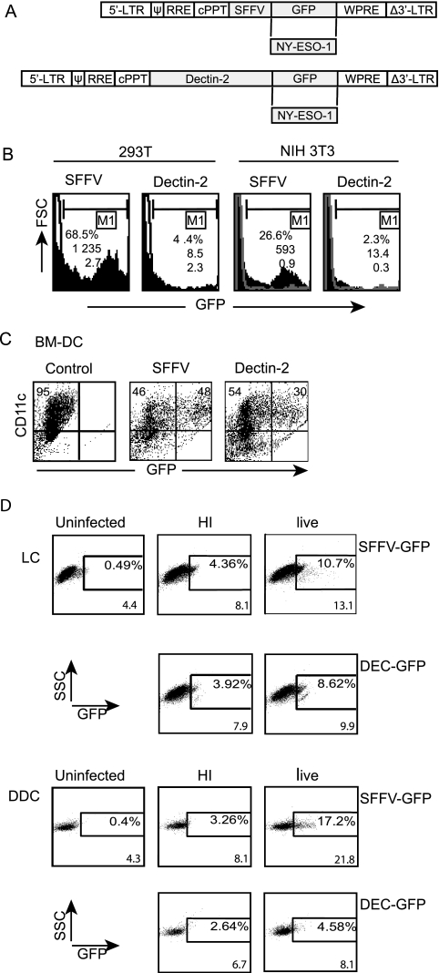
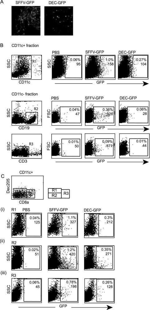
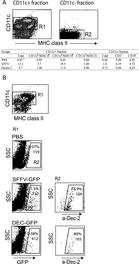
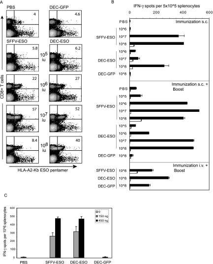
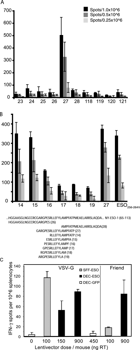
Lentivectors stimulate potent immune responses to antigen transgenes and are being developed as novel genetic vaccines. To improve safety while retaining efficacy, we constructed a lentivector in which transgene expression was restricted to antigen-presenting cells using the mouse dectin-2 gene promoter. This lentivector expressed a green fluorescent protein (GFP) transgene in mouse bone marrow-derived dendritic cell cultures and in human skin-derived Langerhans and dermal dendritic cells. In mice GFP expression was detected in splenic dectin-2(+) cells after intravenous injection and in CD11c(+) dendritic cells in the draining lymph node after subcutaneous injection. A dectin-2 lentivector encoding the human melanoma antigen NY-ESO-1 primed an NY-ESO-1-specific CD8(+) T-cell response in HLA-A2 transgenic mice and stimulated a CD4(+) T-cell response to a newly identified NY-ESO-1 epitope presented by H2 I-A(b). As immunization with the optimal dose of the dectin-2 lentivector was similar to that stimulated by a lentivector containing a strong constitutive viral promoter, targeting antigen expression to dendritic cells can provide a safe and effective vaccine.


Figures

FIG. 1.
FIG. 1.
Lentiviral vectors, transgene expression in vitro. (A) Lentiviral vector (pHRSINCSGW) expressing GFP under the SFFV promoter was used as a control. Experimental vectors were made by replacing SFFV with the dectin-2 gene promoter and GFP with NY-ESO-1 antigen. cPPT, central polypurine tract; WPRE, woodchuck hepatitis virus posttranscriptional regulatory element; LTR, long terminal repeat; RRE, Rev response element. (B) Human 293T cells and murine NIH 3T3 cells were transduced (MOI of 1) with SFFV-GFP or DEC-GFP lentivectors, and GFP expression was analyzed by FACS 72 h postinfection (solid profiles); nontransduced cells are shown as empty profiles. Numbers indicate percentage of transduced cells, MFI values, and the number of copies of transgene per cell (as determined by TaqMan PCR). (C) Bone marrow-derived DC (BM-DC) were transduced with SFFV-GFP or DEC-GFP lentivectors (MOI of 10), stained with anti-CD11c-PE antibody on day 6 postinfection, and then analyzed for GFP expression by FACS. Cell surface staining shows that 95% of the cell population was CD11c+. Numbers in the upper right quadrants indicate the percentage of GFP-expressing cells among the CD11c+ cells, with MFI values for GFP as 4,161 and 2,256 for the SFFV and dectin-2 promoters, respectively. This is a representative result out of four experiments performed. (D) Lentiviral transduction of primary human LC and dDC. Both cell populations were characterized by surface staining with cell-specific makers. The LC were CD45+ DR+ CD1a+ Langerin+ CD14 and the dDCs were CD45+ DR+ CD1alow CD14 (not shown). Only DR staining is shown for the sake of simplicity. Cells were transduced with SFFV-GFP or DEC-GFP lentivectors (MOI of 60), and GFP expression was detected by FACS after gating on forward-scatter high HLA-DR+ cells in the preparation. Comparison of uninfected controls and cells exposed to heat-inactivated virus (HI) and live virus are shown. Numbers indicate percentages of transduced cells and MFI values. This experiment was performed 3 times with similar results. FSC, forward scatter; SSC, side scatter.
FIG. 2.
FIG. 2.
Transgene expression following subcutaneous injection. (A). The epidermal sheet obtained from mouse ear skin was treated in vitro with lentivectors, and skin was analyzed for GFP expression 48 h later. (B). Transgene expression in LN. Mice were injected subcutaneously with PBS (control) or SFFV-GFP or DEC-GFP lentivector. Five days later, LNs were collected, cells were pooled (n = 3 mice/group), and CD11c+- and CD11c-enriched cell fractions were obtained using CD11c microbeads. Cells from each fraction were analyzed for GFP expression by FACS after staining with specific antibodies. The CD11c+ fraction was stained with anti-CD11c antibody and region R1 was established. GFP+ cells in the CD11c fraction were then analyzed within the R1 region. Cells from the CD11c fraction were stained for B- and T-cell markers (anti-CD19 or anti-CD3 antibodies, respectively), and analysis of GFP+ cells in each group was performed after gating in the corresponding population. (C). LN DC were costained with anti-CD11c, anti-DEC205, and anti-CD8α antibodies, and three DC subsets were identified, as shown in the diagram. Analysis of GFP expression within each region was then performed. Numbers indicate the percentages of transduced cells and the MFI values. At least 30,000 events per sample were analyzed. This experiment was repeated twice with similar results. SSC, side scatter.
FIG. 3.
FIG. 3.
Transgene expression in spleen following intravenous injection. Mice were injected intravenously with PBS (control) or SFFV-GFP or DEC-GFP lentivector. Spleen cells were collected and pooled (10 days postinfection; n = 2 mice/group), and CD11c+ cells were isolated using CD11c microbeads. Transgene expression in both CD11c+- and CD11c-enriched cell fractions was analyzed by FACS after costaining for cell surface-specific markers as indicated. The CD11c+ fraction contains ≥75% CD11c+ cells, the CD11c fraction is ≥96% CD11c. The table shows the percentage of GFP+ cells in three subpopulations of cells within the CD11c+ cell fraction, according to their CD11c and MHC-II expression levels (as defined in panel A) and within the CD11C fraction after costaining with anti-CD19 and anti-CD3 antibodies. The CD11clo/MHC-IIhi population includes autofluorescent and F4/80+ cells, consistent with the description for spleen macrophages. (B) Surface expression of dectin-2 protein in GFP+ cells in the CD11c+ fraction. Cells were stained with CD11c, MHC-II, and dectin-2 antibodies, and region R1 was established according to CD11c and MHC-II+ staining. GFP+ cells within R1 were then gated (R2), and the percentage of dectin-2-expressing cells in R2 (i.e., GFP+) was determined. Numbers represent the percentage of positive cells for the marker expressed in the x axis and the MFI value. Overall, dectin-2+ cells in the CD11c+ fraction in all three groups corresponded to ≤7% of the CD11c+ population. The data represent one out of five experiments performed with similar results. SSC, side scatter.
FIG. 4.
FIG. 4.
NY-ESO-specific CD8+ T-cell responses after immunization with lentivectors. (A and B) HHD mice were immunized subcutaneously or intravenously with PBS, DEC-GFP, SFFV-ESO, or DEC-ESO VSV-G lentivectors. Lentivector doses per mouse are indicated. Some mice received an intravenous boost with ESO-vaccinia virus. (A) CD8+ T-cell response measured by pentamers (day 8 after boost) in the blood of mice that were immunized subcutaneously and boosted. Numbers indicate the percentages of T cells responding to the HLA-A201Kb-ESO-157 pentamers within the CD8+ population. Data are from one mouse out of two per group. (B) T cells were tested in an ELISPOT IFN-γ assay 12 days after immunization or boost. Spleen cells were pooled (n = 2/group), and incubated in duplicates, either with the HLA-A021-restricted ESO-1157-165 peptide (black bars) or medium only (white bars). Standard error bars represent the mean count from duplicate wells. One representative experiment out of three is shown. s.c., subcutaneous; i.v., intravenous. (C) HHD mice were immunized subcutaneously with Friend-pseudotyped lentivectors carrying the ESO peptide, and spleen cells were tested in an ELISPOT IFN-γ assay 12 days later. Spleen cells were pooled (n = 3/group), and incubated with the HLA-A021-restricted ESO-1157-165 peptide. Values are the means ± standard error of duplicate wells after subtraction of background values of cells incubated with medium only.
FIG. 5.
FIG. 5.
CD4+ response to NY-ESO-1 in HLA-A2 transgenic mice. (A) Splenocytes from HHD mice immunized with a DNA vaccine encoding full-length NY-ESO-1 were screened in an ELISPOT IFN-γ recall assay with overlapping 21-mer peptides. Values are the number of spots per the indicated number of splenocytes. (B) Splenocytes from HHD mice immunized with a DNA vaccine encoding full-length NY-ESO-1 were screened in an ELISPOT IFN-γ recall assay with overlapping 14-mer peptides. Numbers along the x axis represent the peptides, whose sequences are given below the graph. Numbers along the x axis represent the peptides; the sequences of critical peptides are below the graph. (C) HHD mice were immunized subcutaneously with PBS or the indicated dose of VSV-G or Friend-pseudoptyped lentivectors, and 12 days later cells were tested in an ELISPOT IFN-γ assay. Spleen cells were pooled (n = 2/group) and incubated in duplicate in either the presence or absence of the ESO-186-99 peptide (peptide 14). Values are the means ± standard error of duplicate wells after subtraction of background values of cells incubated with medium only.

Similar articles

Cited by

References

    1. Allan, R. S., C. M. Smith, G. T. Belz, A. L. van Lint, L. M. Wakim, W. R. Heath, and F. R. Carbone. 2003. Epidermal viral immunity induced by CD8α+ dendritic cells but not by Langerhans cells. Science 3011925-1928. - PubMed
    1. Allan, R. S., J. Waithman, S. Bedoui, C. M. Jones, J. A. Villadangos, Y. Zhan, A. M. Lew, K. Shortman, W. R. Heath, and F. R. Carbone. 2006. Migratory dendritic cells transfer antigen to a lymph node-resident dendritic cell population for efficient CTL priming. Immunity 25153-162. - PubMed
    1. Aragane, Y., A. Maeda, A. Schwarz, T. Tezuka, K. Ariizumi, and T. Schwarz. 2003. Involvement of dectin-2 in ultraviolet radiation-induced tolerance. J. Immunol. 1713801-3807. - PubMed
    1. Ariizumi, K., G. L. Shen, S. Shikano, R. Ritter, 3rd, P. Zukas, D. Edelbaum, A. Morita, and A. Takashima. 2000. Cloning of a second dendritic cell-associated C-type lectin (dectin-2) and its alternatively spliced isoforms. J. Biol. Chem. 27511957-11963. - PubMed
    1. Belz, G. T., K. Shortman, M. J. Bevan, and W. R. Heath. 2005. CD8α+ dendritic cells selectively present MHC class I-restricted noncytolytic viral and intracellular bacterial antigens in vivo. J. Immunol. 175196-200. - PMC - PubMed

Publication types

MeSH terms