Comparative Study
. 2007 Oct 24;27(43):11641-50.
doi: 10.1523/JNEUROSCI.2457-07.2007.

A role of the transient receptor potential domain of vanilloid receptor I in channel gating

Affiliations
Comparative Study

A role of the transient receptor potential domain of vanilloid receptor I in channel gating

Nuria García-Sanz et al. J Neurosci. .

Abstract

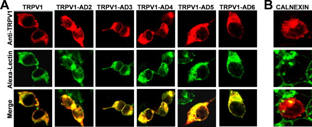
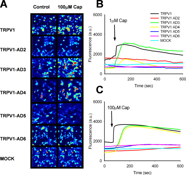
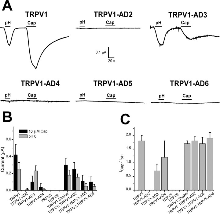
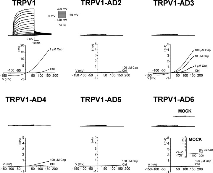
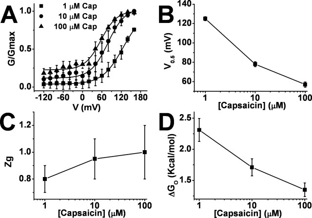
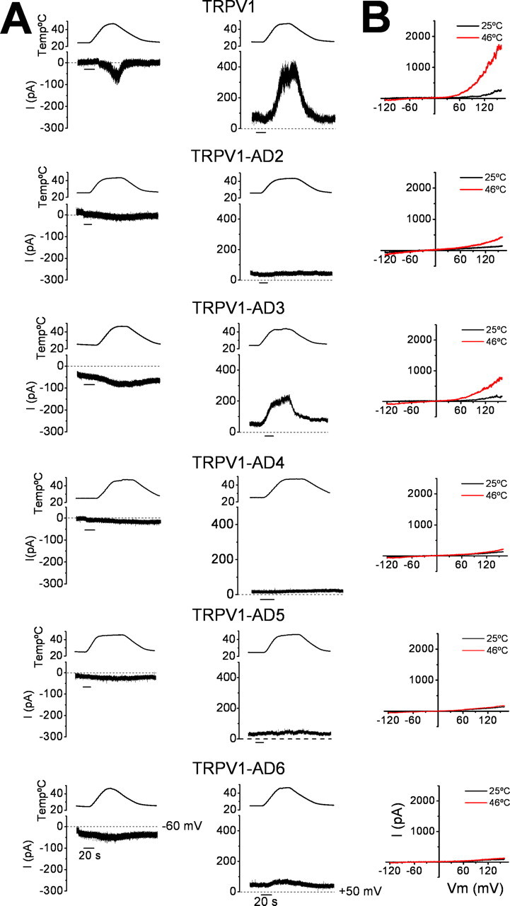
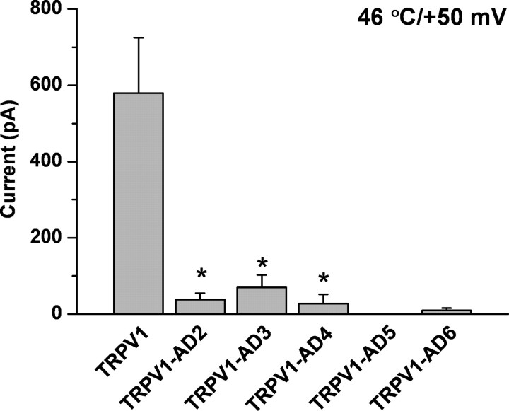
Transient receptor potential vanilloid receptor subtype 1 (TRPV1) is an ionotropic receptor activated by temperature and chemical stimuli. The C-terminal region that is adjacent to the channel gate, recognized as the TRP domain, is a molecular determinant of receptor assembly. However, the role of this intracellular domain in channel function remains elusive. Here, we show that replacement of the TRP domain of TRPV1 with the cognate region of TRPV channels (TRPV2-TRPV6) did not affect receptor assembly and trafficking to the cell surface, although those receptors containing the TRP domain of the distantly related TRPV5 and TRPV6 did not display ion channel activity. Notably, functional chimeras exhibited an impaired sensitivity to the activating stimuli, consistent with a significant contribution of this protein domain to channel function. At variance with TRPV1, voltage-dependent gating of chimeric channels could not be detected in the absence of capsaicin and/or heat. Biophysical analysis of functional chimeras revealed that the TRP domain appears to act as a molecular determinant of the activation energy of channel gating. Together, these findings uncover a role of the TRP domain in intersubunit interactions near the channel gate that contribute to the coupling of stimulus sensing to channel opening.


Figures

Figure 1.
Figure 1.
The TRP domain is conserved among TRPV channels. A, Amino acid sequence alignment of the TRP domain of TRPV channels. The “a” and “d” positions of the predicted coiled coil are boxed. B, Wheel representation of the TRP domain of TRPV1; in red are denoted the positions of a coiled coil. C, Diagram representing the design of TRPV1 chimeras in which the TRP domain (denoted as the AD) was replaced with the cognate of TRPV2 (TRPV1–AD2), TRPV3 (TRPV1–AD3), TRPV4 (TRPV1–AD4), TRPV5 (TRPV1–AD5), and TRPV6 (TRPV1–AD6). CC, Coiled coil score obtained in ExPASy Proteomic server (http://expasy.org/).
Figure 2.
Figure 2.
TRPV1 chimeras are expressed at the cell surface of HEK293 cells. A, Confocal microscopy images of HEK293 cells transfected with TRPV1 and chimeric subunits sequentially exposed to a fluorescently labeled wheat agglutinin (Alexa-Lectin) and to a specific anti-TRPV1 antibody against its N terminus. B, Confocal images of untransfected HEK293 cells sequentially exposed to the fluorescent-lectin and anti-calnexin antibody, a selective marker of the endoplasmic reticulum. Fixed, intact cells were incubated with the fluorescent lectin and thereafter were permeabilized with detergent and incubated with the anti-TRPV1 or the anti-calnexin antibodies.
Figure 3.
Figure 3.
TRPV1–AD3 and TRPV1–AD4 chimeras display capsaicin-evoked Ca+2 influx. A, Ca2+ images showing the intracellular rise in Ca+ evoked by 100 μm capsaicin from HEK293 cells expressing the different TRPV1 species or mock-transfected cells. B, C, Change in Ca2+-dependent fluorescence of TRPV1-transfected and mock cells as a function of time, before and after the exposure to 1 and 100 μm for 10 s at 20–22°C, respectively. Representative images and traces of 50 cells measured in n = 3 different experiments. Traces represent that change in fluorescence per cell. Cells were loaded with Fluo-4 AM to record intracellular calcium signals and activated as indicated. The extracellular concentration of Ca2+ was 2 mm. The arrows indicate the onset of capsaicin exposure.
Figure 4.
Figure 4.
Expression of chimeric channels in Xenopus oocytes. A, Ionic currents activated by pH 6.0 and 10 μm capsaicin from amphibian oocytes injected with the cRNA encoding TRPV1 and chimeric channels. Oocytes were held at −60 mV. Ionic currents were measured 48–72 h after cRNA injection in Mg2+–Ringer's solution at 20°C. Current traces are representative of n ≥ 3 oocytes. B, Mean amplitude of ionic currents elicited by pH 6.0 and 10 μm capsaicin. C, ICap/IpH ratio for TRPV1 and chimeric channels calculated form the recordings shown in A. Data are shown as mean ± SD, with number of oocytes of n ≥ 3.
Figure 5.
Figure 5.
Voltage-dependent gating of TRPV1 and chimeras expressed in HEK293 cells. Representative family of whole-cell currents elicited with a voltage protocol consisting of 50 ms depolarizing pulses from −120 up to 300 mV in steps of 20 mV. I–V relationships evoked at increasing concentrations of capsaicin are shown underneath the ionic currents. Ramps consisted of a voltage step of 300 ms from the holding potential of 0 to −120 mV, followed by 350 ms linear ramp up to 160 mV. Representative current traces from n ≥ 6 cells are shown. Whole-cell currents were obtained in symmetrical 150 mm NaCl, from a holding potential of 0 mV. Ctrl, I–V curves obtained in the absence of capsaicin.
Figure 6.
Figure 6.
Capsaicin-evoked ionic currents from TRPV1–AD3 chimeras. A, Representative family of whole-cell currents elicited with a voltage protocol consisting of 100 ms depolarizing pulses from −120 up to 160 mV in steps of 20 mV for TRPV1–AD3 at increasing concentrations of capsaicin. B, Current densities of TRPV1 and TRPV1–AD3 evoked in the presence of capsaicin at −60 and 160 mV. Ionic currents from TRPV1–AD3 were activated by 100 μm capsaicin, whereas those of TRPV1 were evoked by 1 μm. Higher capsaicin concentrations could not be used for TRPV1 because the large currents evoked at positive potentials could not be clamped. C, Dose–response relationships for capsaicin activation of TRPV1 and TRPV1–AD3 chimera. Channel activity was elicited at a holding potential of −60 mV. Solid lines depict the theoretical fits to a Michaelis–Menten binding isotherm (Garcia-Martinez et al. 2006). The EC50 values for capsaicin were 0.9 ± 0.2 μm for TRPV1 and 10 ± 2 μm for TRPV1–AD3. The Hill coefficients were 1.8 ± 0.2 for TRPV1 and 1.4 ± 0.2 for TRPV1–AD3. Responses were normalized to the maximal capsaicin-evoked ionic current inferred from the fit of the dose–response curves depicting the total ionic current recorded at increasing concentrations of capsaicin. Each point represents the mean ± SD, with n = 4. ctrl, I–V curves obtained in the absence of capsaicin.
Figure 7.
Figure 7.
Voltage-dependent properties of TRPV1–AD3 chimera. A, G–V relationships of TRPV1–AD3 at increasing concentrations of capsaicin (1–100 μm). The G–V curves were obtained by converting the maximal current values from a voltage step protocol shown in Figure 6A to conductance using the relation G = I/(VVR), where G is the conductance, I is the peak current, V is the command pulse potential, and VR is the reversal potential of the ionic current obtained form the I–V curves. Solid lines depict the best fit to a Boltzmann distribution. B, C, Variation of the V0.5 (B) and zg (C) of TRPV1–AD3 as a function of the capsaicin concentration, respectively. The apparent gating valence of the activation process obtained using zg = 25.69 mV/an, where an is the slope of the G–V curves. V0.5 and an values were obtained from the fit of G–V relationships depicted in A to a Boltzmann distribution. D, Variation of the free energy (ΔGo) as a function of the capsaicin concentration. The ΔGo was obtained from ΔGo = zgFV0.5, where F is the faraday constant (0.023 kcal · mol−1 · mV−1). Data are given as mean ± SD, with n ≥ 5 cells. *p < 0.05 obtained with the Student's t test.
Figure 8.
Figure 8.
Heat-dependent gating of TRPV1 and chimeric channels. A, Traces represent the ionic currents evoked by heat ramps (depicted on top, 0.3°C/s) from cells held at −60 mV (left) and +50 mV (right). B, I–V curves obtained at 25°C and 46°C for wild-type and chimeric channels. Cells were depolarized from −120 to +160 mV in 350 ms with a linear ramp. Traces are representative of n ≥ 3 cells.
Figure 9.
Figure 9.
Temperature-dependent properties of wild-type and TRPV1–AD3 chimera. Mean amplitude of the ionic currents evoked by a heat ramp from 25°C to 46°C recorded at +50 mV and 46°C for TRPV1 and chimeric channels. Ionic currents were obtained from the recordings shown in Figure 7. Data are given as mean ± SD, with n ≥ 3 cells. *p < 0.05 obtained with Student's t test.
Figure 10.
Figure 10.
Effect of heat on the capsaicin responses of TRPV1, TRPV1–AD2, TRPV1–AD3, and TRPV1–AD4 chimeras. Mean amplitude of the ionic currents evoked by heat (45°C) and 1 μm capsaicin at 25°C and 45°C for TRPV1, TRPV1–AD2, TRPV1–AD3, and TRPV1–AD4 chimeras. Ionic currents were recorded at +50 mV. Data are given as mean ± SD, with n ≥ 3 cells. *p < 0.05 obtained with the Student's t test.

Similar articles

Cited by

References

    1. Arniges M, Fernandez-Fernandez JM, Albrecht N, Schaefer M, Valverde MA. Human TRPV4 channel splice variants revealed a key role of ankyrin domains in multimerization and trafficking. J Biol Chem. 2006;281:1580–1586. - PubMed
    1. Bandell M, Dubin AE, Petrus MJ, Orth A, Mathur J, Hwang SW, Patapoutian A. High-throughput random mutagenesis screen reveals TRPM8 residues specifically required for activation by menthol. Nat Neurosci. 2006;9:493–500. - PubMed
    1. Brauchi S, Orio P, Latorre R. Clues to understanding cold sensation: thermodynamics and electrophysiological analysis of the cold receptor TRPM8. Proc Natl Acad Sci USA. 2004;101:15494–15499. - PMC - PubMed
    1. Brauchi S, Orta G, Salazar M, Rosenmann E, Latorre R. A hot-sensing cold receptor: C-terminal domain determines thermosensation in transient receptor potential channels. J Neurosci. 2006;26:4835–4840. - PMC - PubMed
    1. Caprini M, Fava M, Valente P, Fernandez-Ballester G, Rapisarda C, Ferroni S, Ferrer-Montiel A. Molecular compatibility of the channel gate and the N terminus of S5 segment for voltage-gated channel activity. J Biol Chem. 2005;280:18253–18264. - PubMed

Publication types

MeSH terms

LinkOut - more resources