Comparative Study
. 2007 Oct 24;27(43):11769-75.
doi: 10.1523/JNEUROSCI.1938-07.2007.

LKB1 regulates neuronal migration and neuronal differentiation in the developing neocortex through centrosomal positioning

Affiliations
Comparative Study

LKB1 regulates neuronal migration and neuronal differentiation in the developing neocortex through centrosomal positioning

Naoyuki Asada et al. J Neurosci. .

Abstract

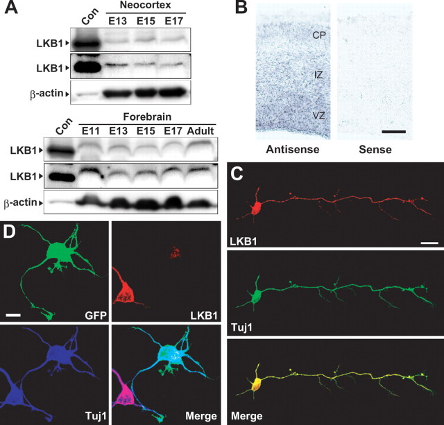
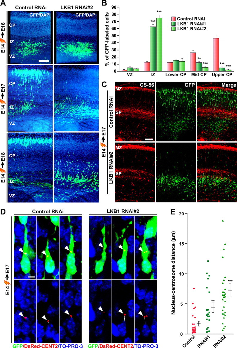
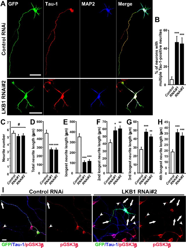
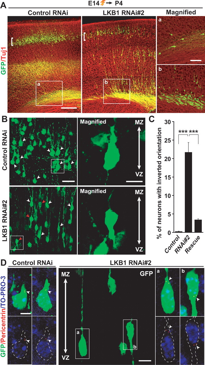
The cerebral cortex is formed through the coordination of highly organized cellular processes such as neuronal migration and neuronal maturation. Polarity establishment of neurons and polarized regulation of the neuronal cytoskeleton are essential for these events. Here we find that LKB1, the closest homolog of the Caenorhabditis elegans polarity protein Par4, is expressed in the developing neocortex. Knock-down of LKB1 in migrating immature neurons impairs neuronal migration, with alteration of the centrosomal positioning and with uncoupling between the centrosome and nucleus. Furthermore, impairment of LKB1 in differentiating neurons within the cortical plate induces malpositioning of the centrosome at the basal side of the nucleus, instead of the normal apical positioning. This is accompanied with the disruption of axonal and dendritic polarity, resulting in reversed orientation of differentiating neurons. Moreover, LKB1 specifies axon and dendrites identity in vitro. Together, these observations indicate that LKB1 plays a critical role in neuronal migration and neuronal differentiation. Furthermore, we propose that proper neuronal migration and differentiation are intimately coupled to the precise control of the centrosomal positioning/movement directed by LKB1.


Figures

Figure 1.
Figure 1.
Expression of LKB1 in the developing mouse brain. A, Neocortical lysates (60 μg of protein) and forebrain lysates (120 μg of protein) derived from different ages indicated were immunoblotted with anti-LKB1 polyclonal antibody (top panels) and anti-β-actin monoclonal antibody (mAb) (bottom panels) (see supplemental methods, available at www.jneurosci.org as supplemental material). The expression profile of LKB1 was confirmed by reprobing the blots with a different LKB1 antibody (anti-LKB1 mAb) (middle panels). Lysates derived from HEK293T cells transfected with a LKB1-expressing plasmid were used as a positive control (Con). B, In situ hybridization of E14 sections using sense and antisense probes to Lkb1 mRNA (see supplemental methods, available at www.jneurosci.org as supplemental material). Scale bar, 100 μm. C, Neocortical neurons at DIV 3 were immunostained with antibodies against LKB1 (red) and Tuj1 (green). Scale bar, 20 μm. D, Neocortical neurons were transfected with GFP-plasmid and either LKB1 RNAi#2 construct or control RNAi construct (pBS-U6) at DIV 1, fixed at DIV 3, and immunostained with antibodies against GFP (green), LKB1 (red), and Tuj1 (blue). Scale bar, 10 μm.
Figure 2.
Figure 2.
Knock-down of LKB1 impairs neuronal migration. A–C, Either LKB1 RNAi construct or control RNAi construct (pBS-U6) was electroporated into E14 embryos together with the GFP-plasmid, and brains were fixed at E16, E17, and E18 as indicated. A, Brain sections were immunostained with antibody against GFP (green). Nuclei were stained with 4′,6′-diamidino-2-phenylindole (DAPI; blue). Representative images are shown. Scale bar, 100 μm. B, The percentage of GFP-labeled cells in the VZ, IZ, and CP of the E17 brain sections was calculated and plotted as the mean ± SEM (6, 4, and 5 embryos for control, RNAi#1, and RNAi#2, respectively). **p < 0.01, ***p < 0.001 versus control by two-tailed Welch's t test. C, E17 brain sections were immunostained with antibodies against GFP (green) and CS-56 (red). Scale bar, 100 μm. D, LKB1 RNAi construct or control RNAi construct was electroporated into E14 embryos together with plasmids encoding GFP and DsRed2-CentrinII. In the E17 brain sections, both control RNAi-introduced and LKB1 RNAi-introduced neurons located in the upper IZ display a stereotypical migratory morphology with a leading process. Arrowheads indicate the centrosome labeled by DsRed2-CentrinII. Solid lines indicate the shortest distance between the nucleus (blue, TO-PRO-3 iodide) and the centrosome (red), which was measured and plotted as the mean ± SEM (n = 23, 20, and 28 cells for control, RNAi#1, and RNAi#2, respectively) in E. **p < 0.01, ***p < 0.001 versus control by two-tailed Welch's t test. Scale bar, 5 μm.
Figure 3.
Figure 3.
Loss of function of LKB1 disrupts neuronal polarity and axon specification. LKB1 RNAi construct or control RNAi construct (pBS-U6) was transfected into primary cortical neurons together with GFP-plasmid at DIV 2. At DIV 4, cortical neurons were fixed. A, The neocortical neurons were immunostained with antibodies against Tau-1 (red) and MAP2 (blue). Scale bars, 50 μm. B, The ratio of neurons that have multiple Tau-1-positive neurites were quantified and plotted as the mean ± SEM (n = 60, 54, and 54 cells for control, RNAi#1, and RNAi#2, respectively). ***p < 0.001 versus control by χ2 test. C–H, Total neurite number (C) and total neurite length (D) of the control cells and the RNAi-introduced cells with multiple Tau-1-positive neurites were quantified and presented as the mean ± SEM. The length of the neurite indicated in the y-axis (E–H) was measured and presented as the mean ± SEM. #p > 0.8 by one-way ANOVA. *p < 0.05, **p < 0.01, ***p < 0.001 versus control by two-tailed Welch's t test (n = 20, 14, and 14 cells for control, RNAi#1, and RNAi#2, respectively). I, The neocortical neurons were immunostained with phospho-GSK3β (Ser-9) (red) and Tau-1 (blue) antibodies at DIV 5. The tips of the Tau-1-positive neurites of the GFP-positive and GFP-negative neurons are indicated by arrows and arrowheads, respectively. An RNAi#2-transfected neuron (GFP-positive) in the right two panels does not have phospho-GSK3β signal at the tips of any neurites (arrows). Scale bar, 20 μm.
Figure 4.
Figure 4.
Knock-down of LKB1 results in reversal of the orientation of differentiating neurons in the CP. A, E14 embryos were electroporated, and brains were fixed at P4. Brain sections were then immunostained with antibodies against GFP (green) and Tuj1 (red). a, b, High-magnification images of the boxed regions in the left two panels. The majority of GFP-labeled neurons in the RNAi-introduced brains remain in the lower layers of the neocortex, whereas a small population of GFP-labeled cells in the control RNAi-introduced brains is in the similar region. Scale bars, 100 μm (left panels) and 40 μm (right panels). B–D, The morphology of neurons in the CP region indicated by the brackets in A was analyzed in detail. B, White arrowheads indicate GFP-labeled neurons with a primitive dendrite-like neurite oriented apically. Red arrowheads indicate GFP-labeled neurons with a ventricle-directed dendrite-like neurite. Right panels, High-magnification images of cells that are boxed in the left panels. Scale bars, 30 μm (left panels) and 10 μm (right panels). C, Quantification of the ratio of neurons with inverted orientation. Data are presented as the mean ± SEM (n = 1028, 750, 2191 neurons from 3 embryos each for control, RNAi#2 and rescue, respectively). ***p < 0.001 versus control by χ2 test. D, Nuclei were stained with TO-PRO-3 iodide (blue). Centrosomes in GFP-labeled neurons were labeled with anti-pericentrin antibody (red) and are indicated by arrowheads. Small panels on the left represent high-power images of GFP-labeled cells in the CP in the brains electroporated with control RNAi. The middle panel shows a representative image of GFP-labeled cells in the CP in the brains electroporated with LKB1 RNAi#2. GFP-labeled cells with normal orientation (a) and with inverted orientation (b) are boxed. Small panels on the right are high-magnification images of the normal (a) and inverted (b) cells in the middle panel. GFP-labeled cells in the small panels are outlined by dashed lines. Scale bars, 5 μm (left and right panels), and 10 μm (middle panel).

Similar articles

Cited by

References

    1. Barnes AP, Lilley BN, Pan YA, Plummer LJ, Powell AW, Raines AN, Sanes JR, Polleux F. LKB1 and SAD kinases define a pathway required for the polarization of cortical neurons. Cell. 2007;129:549–563. - PubMed
    1. Etienne-Manneville S, Hall A. Cdc42 regulates GSK-3β and adenomatous polyposis coli to control cell polarity. Nature. 2003;421:753–756. - PubMed
    1. Higginbotham H, Tanaka T, Brinkman BC, Gleeson JG. GSK3β and PKCζ function in centrosome localization and process stabilization during Slit-mediated neuronal repolarization. Mol Cell Neurosci. 2006;32:118–132. - PubMed
    1. Jiang H, Guo W, Liang X, Rao Y. Both the establishment and the maintenance of neuronal polarity require active mechanisms: critical roles of GSK-3β and its upstream regulators. Cell. 2005;120:123–135. - PubMed
    1. Kemphues KJ, Priess JR, Morton DG, Cheng NS. Identification of genes required for cytoplasmic localization in early C. elegans embryos. Cell. 1988;52:311–320. - PubMed

Publication types

Substances

LinkOut - more resources