. 2008 Jan;19(1):95-104.
doi: 10.1091/mbc.e07-06-0565. Epub 2007 Oct 24.

The Mitogen-activated protein kinase p38 links Shiga Toxin-dependent signaling and trafficking

Affiliations

The Mitogen-activated protein kinase p38 links Shiga Toxin-dependent signaling and trafficking

Sébastien Wälchli et al. Mol Biol Cell. 2008 Jan.

Abstract

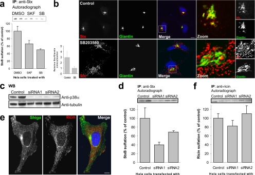
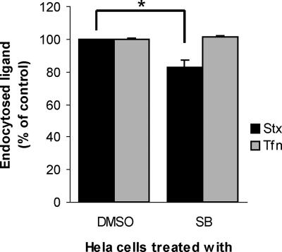
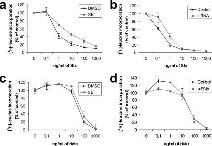
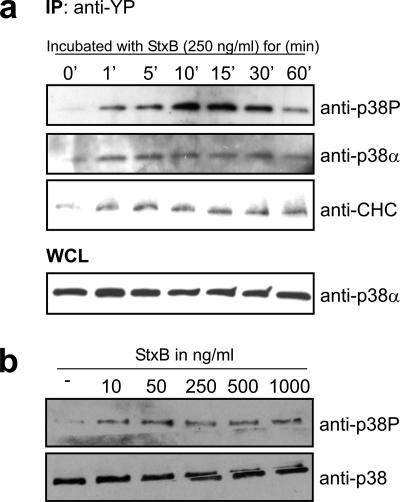
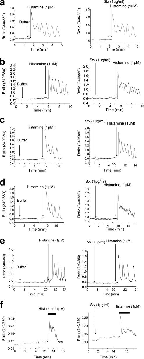
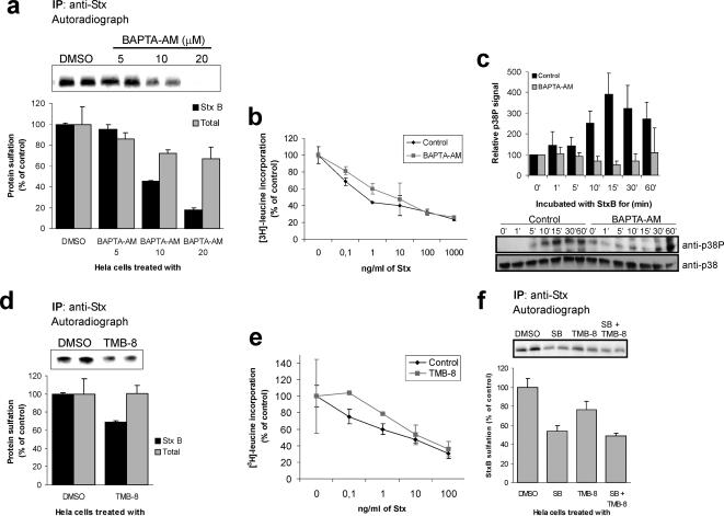
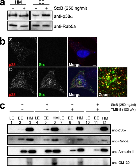
Shiga toxin (Stx) binds to the cell, and it is transported via endosomes and the Golgi apparatus to the endoplasmic reticulum and cytosol, where it exerts its toxic effect. We have recently shown that Stx activates the tyrosine kinase Syk, which in turn induces clathrin phosphorylation and up-regulates Stx uptake. Here, we show that toxin-induced signaling can also regulate another step in intracellular Stx transport. We demonstrate that transport of Stx to the Golgi apparatus is dependent on the mitogen-activated protein kinase p38. Treatment of cells with chemical inhibitors or small interfering RNA targeting p38 inhibited Stx transport to the Golgi and reduced Stx toxicity. This p38 dependence is specific to Stx, because transport of the related toxin ricin was not affected by p38 inhibition. Stx rapidly activated p38, and recruited it to early endosomes in a Ca(2+)-dependent manner. Furthermore, agonist-induced oscillations in cytosolic Ca(2+) levels were inhibited upon Stx stimulation, possibly reflecting Stx-dependent local alterations in cytosolic Ca(2+) levels. Intracellular transport of Stx is Ca(2+) dependent, and we provide evidence that Stx activates a signaling cascade involving cross talk between Ca(2+) and p38, to regulate its trafficking to the Golgi apparatus.


Figures

Figure 1.
Figure 1.
p38 inhibition reduces transport of StxB, but not ricin, to the TGN. (a) HeLa cells were incubated with radioactive sulfate for 3 h, and the indicated inhibitors (SKF86002 and SB203580; 30 μM) or the carrier (dimethyl sulfoxide [DMSO]; 0.1% final concentration) were present for the last 30 min. StxB was then added, and the incubation was continued for 45 min. StxB was immunoprecipitated from the lysates, and its degree of sulfation was analyzed by SDS-PAGE and autoradiography. The band intensities were quantified and the average plotted with error bars showing deviations. The experiment was performed three times with duplicates. (b) HeLa cells were incubated with or without SB203580 for 30 min, before addition of and further incubation with 2 μg/ml StxB for 20 min. The cells were then fixed and permeabilized before staining with the indicated antibodies. DRAQ5 was used for nuclear staining. Bar, 10 μm. Colocalization of StxB with the Golgi marker Giantin was quantified using Zeiss LSM Image Browser. Bars represent SD; n = 10. (c) We analyzed 1/100 of the Stx-IP supernatant (from D) for p38α by Western blot. α-Tubulin was used as loading control. (d) As in a, but in this case cells were transfected with 100 nM of the indicated siRNA and incubated for 48 h before StxB-Sulf2 treatment. These experiments were repeated at least three times with duplicates. (e) Cells were incubated for 20 min with 2 μg/ml Stx and 2 μg/ml ricin before fixation and permeabilization. They were then stained with antibodies as indicated. Bar, 10 μm. (f) As in d, but with 90-min incubation of ricin sulf-1 instead of StxB-Sulf2.
Figure 2.
Figure 2.
Effect of p38 inhibition on endocytosis. HeLa cells were incubated with 30 μM SB203580 (SB) or carrier (DMSO; 0.1% final concentration) for 30 min at 37°C before Stx-SS-Biotin (black bars) or transferrin-SS-Biotin (Tfn; gray bars) was added to the medium. The incubation was continued for 20 min. Cells were 2-mercaptoethane sulphonate sodium treated, and internalized Stx- or transferrin-SS-Biotin labeled with Ru(II)-tag either directly (transferrin) or via antibody (Stx) was fished out from cell lysates by streptavidin-coated magnetic beads and measured by electrochemiluminescence. This experiment was repeated three times with duplicates; error bars represent deviations. *p ≤ 0.005, determined by the paired Student's t test.
Figure 3.
Figure 3.
p38 inhibition protects against Stx, but not ricin, cytotoxicity. HeLa cells were treated with 30 μM SB or carrier (DMSO; 0.1% final concentration) for 30 min (a and c) or transfected with p38α siRNA1 or a control siRNA 48 h before the experiment (b and d). After this, the cells were incubated with increasing concentrations of Stx (a and b) or ricin (c and d) for 2.5 h. Protein synthesis was measured by [3H]leucine incorporation. All experiments were repeated at least twice with duplicates. Error bars represent standard deviations.
Figure 4.
Figure 4.
StxB is able to activate p38α upon binding. (a) HeLa cells were starved for 2 h in HEPES medium before incubation with 250 ng/ml StxB for the indicated times. Cells were lysed, and cell lysate was passed through an anti-YP column. The eluate was then analyzed by Western immunoblotting with the indicated antibodies. One percent of the WCL was analyzed by SDS-PAGE and Western immunoblot to serve as control of equal loading on the column. (b) HeLa cells were incubated with increasing concentrations of StxB for 5 min at 37°C. Lysates were prepared and run for Western blot analysis by using the indicated antibodies. These experiments were repeated three times.
Figure 5.
Figure 5.
Stx inhibits cytosolic Ca2+ oscillations. Cytosolic Ca2+ was measured in HeLa cells, loaded with the calcium probe Fura-2AM. Stx (1 μg/ml) was added to the medium, as indicated, after initiation of Ca2+ measurements. Histamine (1 μM) was added 20 s (a), 4 min (b), 10 min (c), or 15 min (d) after Stx addition. For c and d, the traces are cut between 5 and 10 or 15 min, respectively. The left traces represent cells without toxin. Traces show the Fura-2AM fluorescent ratio (340 nm/380 nm). (e) After Fura-2AM loading, HeLa cells were incubated at 18°C for measurements. Stx was added as indicated. Histamine (1 μM) was added 20 min after the Stx addition. Traces presented are cut between 5 and 20 min. (f) Cells were pretreated with 2 μM SB203580 for 30 min. The cells were then loaded with Fura-2AM. Stx was added 2 min after the measurements started. Histamine (1 μM) was added 15 min after the Stx addition. Each trace is a representative Ca2+ response for 18–30 individual cells per experimental condition.
Figure 6.
Figure 6.
StxB transport to the TGN is sensitive to Ca2+ variations. (a) HeLa cells were incubated with BAPTA-AM at the indicated concentrations or the carrier (DMSO; 0.1% final concentration) for 30 min before incubation with StxB for 45 min and lysis of the cells. StxB was immunoprecipitated from the lysates, and its degree of sulfation analyzed by SDS-PAGE and autoradiography. The band intensities were calculated and plotted as average of parallels. Total cellular proteins sulfation was measured after TCA precipitation and plotted relative to the control. This experiment was performed twice with duplicates; error bars show deviations. (b) Cells were incubated with or without 10 μM BAPTA-AM for 30 min before addition of Stx. The experiment was then performed as described in Figure 3. This experiment was performed twice with duplicates; error bars show deviations. (c) Cells were serum starved for 1 h, and then they were treated with or without 10 μM BAPTA-AM for 30 min before StxB was added. Cell lysates were separated by SDS-PAGE and analyzed by Western blot. Quantification of three separate experiments is shown in the histogram. (d) Cells were incubated with 100 μM TMB-8 or the carrier (DMSO; 0.1% final concentration) for 30 min before incubation with StxB-Sulf2 for 45 min. The experiment was then carried out as described in A. (e) Cells were incubated with or without 100 μM TMB-8 for 30 min before addition of Stx. The experiment was then performed as described in Figure 3. This experiment was repeated three times with duplicates. Error bars show deviations. (f) As in d, but in this assay cells were also treated or not with 30 μM SB203580 before addition of StxB-Sulf2. This experiment was repeated twice with duplicates. Error bars show deviations.
Figure 7.
Figure 7.
p38 is recruited to early endosomes by a Ca2+-dependent mechanism upon Stx binding. (a) HEp2 cells were starved for 1 h before incubation with 250 ng/ml StxB for 20 min. Endosome fractions were prepared as described in Materials and Methods, and equal amounts of proteins were separated by SDS-PAGE and analyzed by Western immunoblotting with the indicated antibodies. (b) HeLa cells were incubated with 2 μg/ml StxB for 0 (top) or 20 min (bottom) before fixation and permeabilization. Antibodies against p38 and Stx were used. Bar, 5 μm. (c) As in a, but cells were treated with or without TMB-8 for 30 min before stimulation with 250 ng/ml StxB. Equal amounts of all fractions were separated by SDS-PAGE and analyzed by Western blot.

Similar articles

Cited by

References

    1. Aniento F., Gu F., Parton R. G., Gruenberg J. An endosomal beta COP is involved in the pH-dependent formation of transport vesicles destined for late endosomes. J. Cell Biol. 1996;133:29–41. - PMC - PubMed
    1. Bencherif M., Eisenhour C. M., Prince R. J., Lippiello P. M., Lukas R. J. The “calcium antagonist” TMB-8 [3,4,5-trimethoxybenzoic acid 8- (diethylamino)octyl ester] is a potent, non-competitive, functional antagonist at diverse nicotinic acetylcholine receptor subtypes. J. Pharmacol. Exp. Ther. 1995;275:1418–1426. - PubMed
    1. Cameron P., Smith S. J., Giembycz M. A., Rotondo D., Plevin R. Verotoxin activates mitogen-activated protein kinase in human peripheral blood monocytes: role in apoptosis and proinflammatory cytokine release. Br. J. Pharmacol. 2003;140:1320–1330. - PMC - PubMed
    1. Cavalli V., Vilbois F., Corti M., Marcote M. J., Tamura K., Karin M., Arkinstall S., Gruenberg J. The stress-induced MAP kinase p38 regulates endocytic trafficking via the GDI:Rab5 complex. Mol. Cell. 2001;7:421–432. - PubMed
    1. Chao T. S., Byron K. L., Lee K. M., Villereal M., Rosner M. R. Activation of MAP kinases by calcium-dependent and calcium-independent pathways. Stimulation by thapsigargin and epidermal growth factor. J. Biol. Chem. 1992;267:19876–19883. - PubMed

Publication types

MeSH terms

LinkOut - more resources