. 2008 Jan;19(1):51-64.
doi: 10.1091/mbc.e07-05-0465. Epub 2007 Oct 24.

Mint3/X11gamma is an ADP-ribosylation factor-dependent adaptor that regulates the traffic of the Alzheimer's Precursor protein from the trans-Golgi network

Affiliations

Mint3/X11gamma is an ADP-ribosylation factor-dependent adaptor that regulates the traffic of the Alzheimer's Precursor protein from the trans-Golgi network

Punya Shrivastava-Ranjan et al. Mol Biol Cell. 2008 Jan.

Abstract

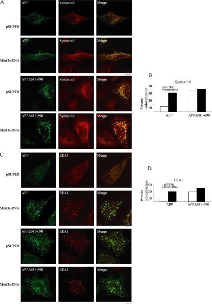
Beta-amyloid peptides (Abeta) are the major component of plaques in brains of Alzheimer's patients, and are they derived from the proteolytic processing of the beta-amyloid precursor protein (APP). The movement of APP between organelles is highly regulated, and it is tightly connected to its processing by secretases. We proposed previously that transport of APP within the cell is mediated in part through its sorting into Mint/X11-containing carriers. To test our hypothesis, we purified APP-containing vesicles from human neuroblastoma SH-SY5Y cells, and we showed that Mint2/3 are specifically enriched and that Mint3 and APP are present in the same vesicles. Increasing cellular APP levels increased the amounts of both APP and Mint3 in purified vesicles. Additional evidence supporting an obligate role for Mint3 in traffic of APP from the trans-Golgi network to the plasma membrane include the observations that depletion of Mint3 by small interference RNA (siRNA) or mutation of the Mint binding domain of APP changes the export route of APP from the basolateral to the endosomal/lysosomal sorting route. Finally, we show that increased expression of Mint3 decreased and siRNA-mediated knockdowns increased the secretion of the neurotoxic beta-amyloid peptide, Abeta(1-40). Together, our data implicate Mint3 activity as a critical determinant of post-Golgi APP traffic.


Figures

Figure 1.
Figure 1.
Mint3 is expressed in neurons of normal human brains. Immunohistochemical staining for Mint3 in normal human brain. (A) In the cerebellar cortex, many Purkinje cells were strongly immunopositive, whereas brain sections in the same region of the cerebellar cortex incubated primary antibody that had been previously incubated with antigen (B) showed negligible staining. (C) In the subiculum, immunoreactive puncta fill the perikarya of pyramidal neurons and extend into apical dendrites. Prior competition with antigen (D) again eliminates the specific staining of Mint3. Bar, 50 μm.
Figure 2.
Figure 2.
Fractionation of traffic components by differential centrifugation of SH-SY5Y homogenates. SH-SY5Y cells were lysed and homogenates prepared before fractionation by differential centrifugation, to generate P1 and P2 pellets and supernatant, S2, as described under Materials and Methods. Equal amounts of protein (15 μg) were resolved by SDS-PAGE and analyzed by immunoblotting with the antibodies indicated on the left of each panel. (A) The distribution of endogenous APP splice variants (APP770 and APP695), Mint 1-3, Fe65, and mannose 6-phosphate receptors (CI-M6PR) is shown. (B) The fractionation of organelle markers for medial-Golgi (GM130), trans-Golgi network (TGN38), endoplasmic reticulum (KDELR), plasma membrane (Na+/K+-ATPase), recycling endosomes (TfR), early endosomes (EEA1), and lysosomes (cathepsin D) in these same fractions is shown. (C) The distribution of vesicle coat proteins AP-1, AP-2, and AP-3 (as determined by immunoblotting with antisera specific to γ-adaptin, α-adaptin, and δ-adaptin, respectively), GGA1-3, β-COP (a component of COPI), clathrin, and the regulatory GTPases Arf3 and Rab6 are shown.
Figure 3.
Figure 3.
Flotation of light vesicles enriched in APP and Mint3 by Optiprep density sedimentation. The pooled fractions from the sucrose velocity centrifugation were brought to 30% Optiprep before under loading in a discontinuous Optiprep gradient, as described under Materials and Methods. Equal volumes (45 μl) from each fraction were analyzed by immunoblot, by using our most sensitive detection method. S2 (20 μg of protein) was used as positive control. Exposure times varied and were the maximum time allowable without background interference, particularly for those antigens showing no reactivity in fractions 3–7 (shown) out of the 16 fractions collected.
Figure 4.
Figure 4.
Increased expression of APP695-Swe resulted in increased APP, Mint3, and Mint2 in the light vesicle fractions. (A) SH-SY5Y cells were infected with lentiviruses that resulted in increased expression of APP695-Swe, Mint3, or both proteins. Equal amounts (15 μg) of total cell homogenates were analyzed for APP and Mint3 expression by immunoblot. (B) Cells were homogenized and equal amounts of protein in S2 were sequentially fractionated by sucrose velocity and Optiprep equilibrium density centrifugation. Equal volumes from fractions 4–10 of the Optiprep gradient were analyzed by immunoblot, with the antibodies shown on the left, with constant exposure times to allow relative quantitation. (C) Fractions from Optiprep gradients of SH-SY5Y or APP695-Swe expressing cells were analyzed for Mint2, as described in B. (D) Enrichment of APP, Mint3, and Mint2 in light vesicles was visualized by comparing equal amounts of protein (1.1 μg) from cell homogenate (H), P1, P2, or fraction 6 from an Optiprep gradient (V6). (E) SH-SY5Y cells expressing APP695-Swe were incubated at 19.5°C for 4 h to block protein exit from the TGN (top; 0 min) or cells were returned to 37°C for 15 min (bottom) before homogenization and light vesicle preparation. Equal volumes from fractions 3–8 of the Optiprep gradients were analyzed by immunoblot, with the antibodies shown on the right and exposure times were constant between cell treatments to allow relative quantitation.
Figure 5.
Figure 5.
APP and Mint3 are present in the same vesicles. Light vesicles (fractions 4–6) from an Optiprep gradient were purified on magnetic beads by using antibodies specific to either the myc epitope (A; as negative control), Mint3 (B), or the C terminus of APP (C) before being analyzed by transmission electron microscopy, as described under Materials and Methods. Representative images are shown of vesicles bound to the surface of the magnetic beads. (D) Vesicles were immunoisolated using Mint3 monoclonal antibodies and were then sequentially incubated with the rabbit polyclonal APP C-terminal antibody and goat anti-rabbit IgG conjugated to 10-nm gold particles before fixation and analysis by electron microscopy, as described under Materials and Methods. (E) Gold-decorated vesicles bound to magnetic beads (from D) are shown enlarged. Figures are representative of three independent experiments. Bar, 100 nm.
Figure 6.
Figure 6.
Decreased expression of Mint3 or mutation of the Mint3 binding domain causes missorting of APP. (A) HeLa cells were transfected with empty vector or either of two different Mint3 siRNA plasmids. The level of expression of Mint3 is shown on day 3 after transfection, as assessed by immunoblotting using antibodies to Mint3 or α-tubulin, which served as a loading control. (B–E) HeLa cells were transfected with empty vector or Mint3 siRNA plasmid and again 2 days later with plasmids driving expression of APP or APPΔ681-690. Analyses were performed 18 h later. (B) Cell lysates were prepared, and equal amounts of total cell protein (25 μg) were resolved in SDS gels, and immunoblotting was performed using antibodies to APP or Mint3. (C) The temperature block was imposed for 4 h before switching to 37°C for 0 or 15 min, as described under Materials and Methods. Cells were then fixed and labeled with antibodies specific to APP for confocal imaging. All images were captured at the same gain and exposure times. Bar, 10 μm. (D) The percentages of cells displaying APP staining in only small puncta (<0.5 μm; empty bars), only a few enlarged puncta (1–4 puncta >0.5 μm; gray bars), or many enlarged puncta (>4 puncta >0.5 μm; black bars) were quantified (APP: pSUPER, n = 142; and Mint3-siRNA, n = 127; APPΔ681-690: pSUPER, n = 138; and Mint3-siRNA, n = 154). Results shown are the average of two independent experiments. (E) Cells were either fixed before temperature block (steady state) or after the block (0 min) or with varying times shown after release from the block, before fixing and staining for APP. Note that steady-state cells do not contain enlarged puncta in either condition and that cells have returned to steady-state staining by 90 min after removal of the block.
Figure 7.
Figure 7.
Decreased expression of Mint3 caused increased APP localization to enlarged endosomal structures. (A–D) Cells were transfected with siRNA and APP or APPΔ681-690 expression plasmids, before imposition of the temperature block and 15-min release, as described in the legend to Figure 6, before fixing and staining for APP and syntaxin 6 (A) or EEA1 (C). Colocalization of APP (green, top two rows of panels) or of APPΔ681–690 (green, bottom two rows) with syntaxin 6 (A; red) or EEA1 (C; red) is dramatically increased (facing page). in cells depleted of Mint3 or with APP mutated in the Mint binding domain, as seen by overlapping (yellow) pixels in the merged image. Quantification of APP and syntaxin 6 (B) or APP and EEA1 (D) colocalization was performed using MetaMorph software, as described under Materials and Methods. Note that colocalization of APPΔ681-690 with both syntaxin 6 or EEA1 is higher than in controls and is largely independent of Mint3 expression levels. Empty bars in B and D are for control (empty pSUPER plasmid) and filled bars are for Mint3siRNA-transfected cells. This experiment was repeated twice with similar results and the quantitation shown is from a single experiment. Bar, 10 μm.
Figure 8.
Figure 8.
Mint3 expression levels impact the rate of secretion of Aβ1-40. HEK293 cells were transiently transfected with either empty vector (control) or vectors directing expression of human Mint1, Mint2, or Mint3. The medium was changed the next day and collected for analysis 48 h later. (A) Conditioned media from cells expressing the different Mints were assayed for Aβ1-40, as described under Materials and Methods. Values represent Aβ1-40 levels relative to controls (mean ± 1 SD; n = 3; **p < 0.001). (B) Cells were transfected with equal amounts of the control or Mint3-siRNA plasmids on day 0 and again on day 3 to maximize knockdown of the protein. Media were replaced on day 4 and conditioned media collected on day 6 for determination of Aβ1-40 levels (n = 3; *p < 0.01). (C) Cells were treated as described in B, and the level of expression of Mint3 was determined in cell lysates (20 μg/lane) by immunoblotting, with α-tubulin used as a loading control. Triplicates are shown of control and Mint3-depleted cell lysates.

Similar articles

Cited by

References

    1. Andersen O. M., et al. Neuronal sorting protein-related receptor sorLA/LR11 regulates processing of the amyloid precursor protein. Proc. Natl. Acad. Sci. USA. 2005;102:13461–13466. - PMC - PubMed
    1. Andersen O. M., et al. Molecular dissection of the interaction between amyloid precursor protein and its neuronal trafficking receptor SorLA/LR11. Biochemistry. 2006;45:2618–2628. - PubMed
    1. Ang A. L., Taguchi T., Francis S., Folsch H., Murrells L. J., Pypaert M., Warren G., Mellman I. Recycling endosomes can serve as intermediates during transport from the Golgi to the plasma membrane of MDCK cells. J. Cell Biol. 2004;167:531–543. - PMC - PubMed
    1. Araki Y., Tomita S., Yamaguchi H., Miyagi N., Sumioka A., Kirino Y., Suzuki T. Novel cadherin-related membrane proteins, Alcadeins, enhance the X11-like protein-mediated stabilization of amyloid beta-protein precursor metabolism. J. Biol. Chem. 2003;278:49448–49458. - PubMed
    1. Bagshaw R. D., Pasternak S. H., Mahuran D. J., Callahan J. W. Nicastrin is a resident lysosomal membrane protein. Biochem. Biophys. Res. Commun. 2003;300:615–618. - PubMed

Publication types

MeSH terms