. 2008 Jan;19(1):237-47.
doi: 10.1091/mbc.e07-02-0179. Epub 2007 Oct 24.

Expansion of the nucleoplasmic reticulum requires the coordinated activity of lamins and CTP:phosphocholine cytidylyltransferase alpha

Affiliations

Expansion of the nucleoplasmic reticulum requires the coordinated activity of lamins and CTP:phosphocholine cytidylyltransferase alpha

Karsten Gehrig et al. Mol Biol Cell. 2008 Jan.

Abstract

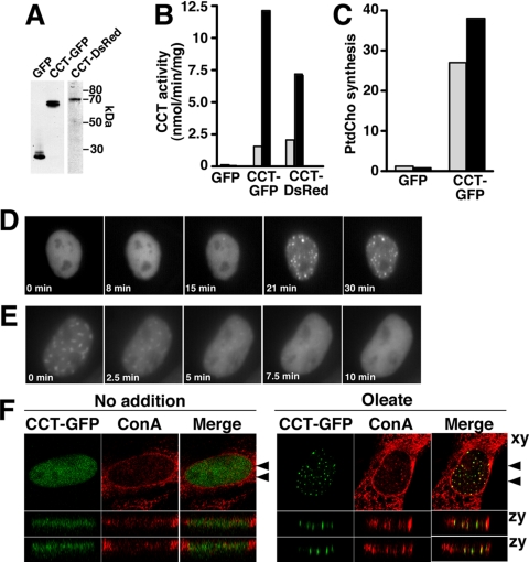
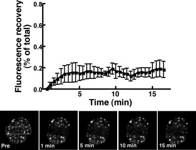
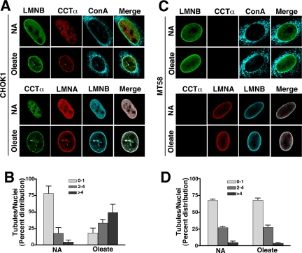
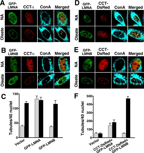
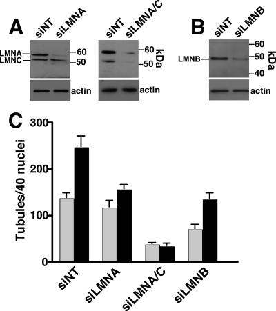
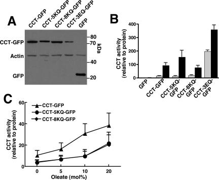
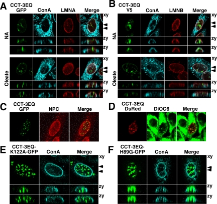
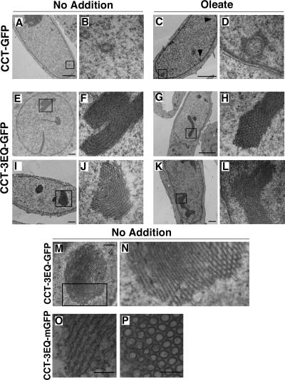
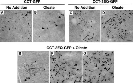
The nucleoplasmic reticulum (NR), a nuclear membrane network implicated in signaling and transport, is formed by the biosynthetic and membrane curvature-inducing properties of the rate-limiting enzyme in phosphatidylcholine synthesis, CTP:phosphocholine cytidylyltransferase (CCT) alpha. The NR is formed by invagination of the nuclear envelope and has an underlying lamina that may contribute to membrane tubule formation or stability. In this study we investigated the role of lamins A and B in NR formation in response to expression and activation of endogenous and fluorescent protein-tagged CCTalpha. Similarly to endogenous CCTalpha, CCT-green fluorescent protein (GFP) reversibly translocated to nuclear tubules projecting from the NE in response to oleate, a lipid promoter of CCT membrane binding. Coexpression and RNA interference experiments revealed that both CCTalpha and lamin A and B were necessary for NR proliferation. Expression of CCT-GFP mutants with compromised membrane-binding affinity produced fewer nuclear tubules, indicating that the membrane-binding function of CCTalpha promotes the expansion of the NR. Proliferation of atypical bundles of nuclear membrane tubules by a CCTalpha mutant that constitutively associated with membranes revealed that expansion of the double-bilayer NR requires the coordinated assembly of an underlying lamin scaffold and induction of membrane curvature by CCTalpha.


Figures

Figure 1.
Figure 1.
CCT-GFP reversibly translocates to NR tubules. (A) GFP, CCT-GFP, or CCT-DsRed were transiently overexpressed in CHO58 cells for 14 h at 33°C before switching to 40°C for 1 h. Total cell lysates (20 μg) were separated by SDS-10%PAGE and immunoblotted using a GFP (CCT-GFP and GFP lanes) or CCTα (CCT-DsRed lane) polyclonal antibody. (B) CCT activity in soluble fractions isolated from CHO58 cells expressing GFP, CCT-GFP, or CCT-DsRed were assayed in the absence (▩) or presence (■) of 1 mM PtdCho/oleate (1:1, mol/mol) vesicles. Results are the mean of duplicates from a representative experiment. (C) [3H]Choline incorporation into PtdCho (DPM/μg cell protein) was quantified in CHO58 cells transiently expressing GFP or CCT-GFP at 40°C. Cells were pulse-labeled in the absence (▩) or presence (■) of 300 μM oleate/BSA for 1 h. Results are the mean of duplicates from a representative experiment. (D) CHO58 cells transiently expressing CCT-GFP at 40°C were exposed to 300 μM oleate/BSA. Images were captured (150–200-ms exposures) at 30-s intervals for up to 30 min using a Photometrics Cascade 512B CCD camera (Woburn, MA) and Metamorph software program (Universal Imaging, West Chester, PA). (E) CHO58 cells were stimulated with oleate/BSA for 20 min as described in D. Medium was then replaced with prewarmed (40°C) oleate-free F12 medium with 0.2% BSA, and images were captured at 30-s intervals. (F) CHO58 cells expressing CCT-GFP were treated with oleate or no addition for 15 min, fixed, and stained with AlexaFluor647-conjugated ConA. Confocal sections (0.2 μm) in the xy plane (top panels) were reconstructed to give two cross-sectional views (indicated by arrows) of the zy plane (lower panels).
Figure 2.
Figure 2.
CCT-GFP associates stably with the NR. CHO58 cells expressing CCT-GFP were treated with oleate/BSA for 20 min as described in the legend to Figure 1D. Photobleaching experiments were performed using Nikon EclipseT2000-E inverted microscope equipped with a 100× oil immersion lens and Nikon Eclipse-D C1 confocal with a 488-nm argon laser. Selected areas of nuclei were photobleached (indicated by the boxed area in the prebleach cell) with the 488-nm laser at full power, and images were captured at 30-s intervals (150–200-ms exposures) for 15–20 min. Fluorescence recovery was calculated using the ratio of fluorescence in the bleached nuclear region/total cell fluorescence at each time point divided by the initial prebleach ratio. Results are the mean and SD of three experiments that examined a total of 12 nuclei each.
Figure 3.
Figure 3.
Formation of a lamin-enriched nucleoplasmic reticulum requires CCTα. (A) CHOK1 cells cultured in DMEM with 5% delipidated FCS received no addition (NA) or 350 μM oleate/BSA for 20 min before fixing and permeabilization with Triton X-100. CCTα was localized with a primary polyclonal antibody and AlexaFluor594- (top panel) or AlexaFluor488-conjugated (bottom panel) secondary antibodies. Lamin B1 was localized with primary and AlexaFluor488 (top panel) or AlexaFluor647 (bottom panel) secondary antibodies. Lamin A/C was visualized with a monoclonal primary antibody and AlexaFluor594 secondary antibody (bottom panel). ER, NE, and NR membranes were visualized with AlexaFluor647-ConA (top panel). (B). The percent distribution of ConA-positive NR tubules/nuclei was quantified in cells treated with oleate or no addition (NA). Results are the mean and SD of 40 nuclei from three separate experiments. (C) CHO58 cells cultured at 40°C were treated with and without oleate and immunostained as described in A. (D) The percent distribution of ConA-positive NR tubules/nuclei was quantified in CHO58 cells. Results are the mean and SD of 40 nuclei from three separate experiments. LMNA, lamin A; LMNB, lamin B1.
Figure 4.
Figure 4.
Lamin A and B1 expression promotes NR proliferation. (A and B) CHOK1 cells were transiently transfected with GFP-lamin A (A) or GFP-lamin B1 (B). Cells then received no addition (NA) or 350 μM oleate/BSA for 20 min at 37°C and were fixed and incubated with AlexaFluor647-ConA. Endogenous CCTα was detected with a primary polyclonal and AlexaFluor594-conjugated secondary antibodies. (C) ConA-positive tubules were quantified in 40 nuclei of control (▩) or oleate-treated (■) cells from three separate experiments. (D and E) CHO58 cells were double-transfected with GFP-lamin A and CCT-DsRed (D) or GFP-Lamin B1 and CCT-DsRed (E) at 37°C, treated with oleate or no addition at 40°C, and incubated with AlexaFluor647-ConA as described above. (F) ConA-positive tubules were quantified in 40 nuclei of control (▩) or oleate-treated (■) cells from three separate experiments.
Figure 5.
Figure 5.
Lamin A/C and B depletion by RNAi prevents NR tubule proliferation. (A) CHOK1 cells were transiently transfected with a nontargeting siRNA (NT) or siRNAs specific for lamin A of lamin A/C. After 48 h, total cell lysates were prepared and immunoblotted with a lamin A/C antibody. Filters were also probed for actin to demonstrate similar protein loading. (B) CHOK1 cells were transfected with an siNT or a lamin B1-specific siRNA, and total cell lysates immunoblotted with a lamin B1-specific antibody. (B) After transient transfection for 48 h with nontargetting, lamin A, or lamin B1 siRNAs, cells were treated with or without 350 μM oleate/BSA in serum-free DMEM for 30 min, fixed and incubated with AlexaFlour488-ConA, and immunostained for lamin A/C and lamin B1. The frequency of NR tubules based on Alexafluor488-ConA staining was quantified in 40 nuclei from untreated (▩) or oleate-treated (■) cells. Results are the mean and SD of three separate experiments. Abbreviations: LMNA, lamin A; LMNA/C, lamin A/C; LMNB, lamin B1.
Figure 6.
Figure 6.
Enzyme activity of GFP-CCTα domain M mutants. (A) Domain M mutants of CCTα and empty vector (GFP) were transiently transfected into CHO58 cells at 37°C for 12 h. Cells were then transferred to 40°C for 30 min, and total cell lysates were prepared and immunoblotted for CCT-GFP fusion proteins with an anti-GFP antibody. Blots were also probed with actin to demonstrate equal protein load. (B) CHO58 cell lysates were assayed for CCT activity in the absence (▩) or presence (■) of 1 mM PtdCho/oleate (1:1, mol/mol) vesicles. Activity was normalized to expression of GFP or CCT-GFP proteins as determined by densitometry. Results are the mean and SD of three separate experiments. (C) Activity of wild type, CCT-5KQ, and CCT-8KQ was assayed using PtdCho vesicles with increasing mol% oleate as described in B. Results are the mean and SD of three experiments.
Figure 7.
Figure 7.
CCTα domain M mutants with reduced membrane affinity do not promote NR proliferation. CHO58 cells transiently expressing (A) CCT-GFP, (B) CCT-5KQ-GFP, or (C) CCT-8KQ-GFP were transferred to 40°C for 1 h and then treated with no addition (NA) or 300 μM oleate/BSA for 15 min. Cells were processed for immunofluorescence and incubated with AlexaFluor647-ConA and with a lamin B1 primary and AlexaFluor594 secondary antibody. (D) In each case, NR tubule frequency was quantified based on AlexaFluor647-ConA staining of control (▩) or oleate-treated (■) cells. Results are the mean and SD of three experiments.
Figure 8.
Figure 8.
Constitutively active, membrane-associated CCT-3EQ promotes nuclear membrane proliferation. (A) CHO58 cells were transiently transfected with CCT-3EQ-GFP at 37°C, transferred to serum-free conditions at 40°C for 1 h, and then treated with addition (NA) or 300 μM oleate/BSA for 15 min at 40°C. Cells were processed for immunofluorescence, incubated with AlexaFluor647-ConA, and immunostained with lamin A/C primary and AlexaFluor594 secondary antibodies. A single 0.5-μm confocal section in the xy plane is shown. Serial confocal sections (15 sections of 0.5 μm each) were used to reconstruct the two images in the zy plane (arrows in xy plane indicate the positions). (B) CHO58 cells were transiently transfected with pcDNA-CCT-3EQ-V5/His and treated as described in A. Fixed cells were incubated with AlexaFluor647-ConA and immunostained with V5 and lamin B1 primary and AlexaFluor-488 or -594 secondary antibodies, respectively. (C) CHO58 cells transiently expressing CCT-3EQ-GFP were fixed and immunostained with nuclear pore complex (NPC)-specific antibody against Nup62 and an AlexaFluor-594 secondary antibody. (D) CHO58 cells transiently expressing CCT-3EQ-DsRed were incubated with DiOC6 to visualize total cellular membranes. (E and F) CHO58 cells were transiently transfected with catalytically dead CCT-3EQ/H89G-GFP or CCT-3EQ/K122A-GFP, treated with 300 μM oleate/BSA for 15 min, stained with AlexaFluor674-ConA, and viewed by serial confocal sections as described in A.
Figure 9.
Figure 9.
CCT-3EQ causes formation of intranuclear stacks of membrane tubules. CHO58 cells were transfected with CCT-GFP or CCT-3EQ-GFP at 37°C, switched to 40°C for 1 h, and treated without (NA) or with 300 μM oleate/BSA for 15 min. Cells were then fixed, embedded, and sectioned for EM analysis. Low-magnification fields are shown in A, C, E, G, I, and K (bar, 2 μm), and corresponding selected high-magnification fields are shown in B, D, F, H, J, and L. (M) A large membrane stack in a CCT-3EQ-GFP–expressing cell (bar, 500 nm). (N) Magnification of selected field from M. (O and P) Longitudinal and cross section of tubule bundles from nuclei of CCT-3EQ-mGFP–expressing cells (bar, 100 nm).
Figure 10.
Figure 10.
CCT-3EQ-GFP is interspersed in membrane tubule stacks. CHO58 cells were transfected with CCT-GFP or CCT-3EQ-GFP and treated with or without oleate as described in the legend to Figure 9. Ultra-thin sections of fixed cells were analyzed by immuno-EM using an anti-GFP polyclonal and goat anti-rabbit secondary conjugated to 5-nm colloidal gold. (A–D) Representative fields from control and oleate-treated cells (bars, 100 nm). Arrows in B indicate NR-like structures. Location of NE is indicated in A and B. (E) Low-magnification of a membrane stack from an oleate-treated CCT-3EQ-GFP–transfected CHO58 cell nuclei (bar, 500 nm). Location of NE is indicated. (F and G) Two selected high-magnification fields from E (bar, 100 nm). Arrows indicate regions of cross section (F) and lengthwise section (G) of membrane stacks.

Similar articles

Cited by

References

    1. Attard G. S., Templer R. H., Smith W. S., Hunt A. N., Jackowski S. Modulation of CTP:phosphocholine cytidylyltransferase by membrane curvature elastic stress. Proc. Natl. Acad. Sci. USA. 2000;97:9032–9036. - PMC - PubMed
    1. Bonne G., et al. Mutations in the gene encoding lamin A/C cause autosomal dominant Emery-Dreifuss muscular dystrophy. Nat. Genet. 1999;21:285–288. - PubMed
    1. Brandt A., et al. Developmental control of nuclear size and shape by Kugelkern and Kurzkern. Curr. Biol. 2006;16:543–552. - PubMed
    1. Broers J. L., Machiels B. M., van Eys G. J., Kuijpers H. J., Manders E. M., van Driel R., Ramaekers F. C. Dynamics of the nuclear lamina as monitored by GFP-tagged A-type lamins. J. Cell Sci. 1999;112(Pt 20):3463–3475. - PubMed
    1. Cornell R. Chemical cross-linking reveals a dimeric structure for CTP:phosphocholine cytidylyltransferase. J. Biol. Chem. 1989;264:9077–9082. - PubMed

Publication types

MeSH terms

Substances

LinkOut - more resources