

Multivariate and univariate analysis of continuous arterial spin labeling perfusion MRI in Alzheimer's disease
- PMID: 17960142
- PMCID: PMC2711077
- DOI: 10.1038/sj.jcbfm.9600570
Multivariate and univariate analysis of continuous arterial spin labeling perfusion MRI in Alzheimer's disease
Abstract
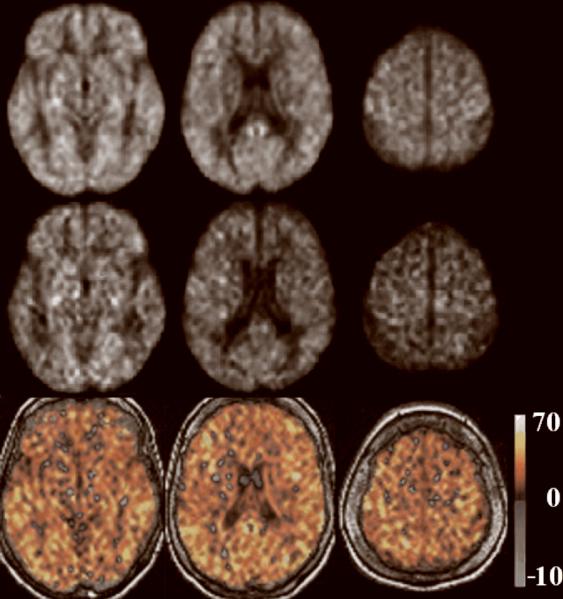
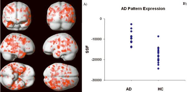
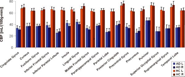
Continuous arterial spin labeling (CASL) magnetic resonance imaging (MRI) was combined with multivariate analysis for detection of an Alzheimer's disease (AD)-related cerebral blood flow (CBF) covariance pattern. Whole-brain resting CBF maps were obtained using spin echo, echo planar imaging (SE-EPI) CASL in patients with mild AD (n=12, age=70.7+/-8.7 years, 7 males, modified Mini-Mental State Examination (mMMS)=38.7/57+/-11.1) and age-matched healthy controls (HC) (n=20; age=72.1+/-6.5 years, 8 males). A covariance pattern for which the mean expression was significantly higher (P<0.0005) in AD than in HC was identified containing posterior cingulate, superior temporal, parahippocampal, and fusiform gyri, as well as thalamus, insula, and hippocampus. The results from this analysis were supplemented with those from the more standard, region of interest (ROI) and voxelwise, univariate techniques. All ROIs (17/hemisphere) showed significant decrease in CBF in AD (P<0.001 for all ROIs, alphacorrected=0.05). The area under the ROC curve for discriminating AD versus HC was 0.97 and 0.94 for covariance pattern and gray matter ROI, respectively. Fewer areas of depressed CBF in AD were detected using voxelwise analysis (corrected, P<0.05). These areas were superior temporal, cingulate, middle temporal, fusiform gyri, as well as inferior parietal lobule and precuneus. When tested on extensive split-half analysis to map out the replicability of both multivariate and univariate approaches, the expression of the pattern from multivariate analysis was superior to that of the univariate.
Figures


References
-
- Alsop DC, Detre JA. Reduced transit-time sensitivity in noninvasive magnetic resonance imaging of human cerebral blood flow. J Cereb Blood Flow Metab. 1996;16:1236–1249. - PubMed
-
- Alsop DC, Detre JA. Multisection cerebral blood flow MR imaging with continuous arterial spin labeling. Radiology. 1998;208:410–416. - PubMed
-
- Alsop DC, Detre JA, Grossman M. Assessment of cerebral blood flow in Alzheimer's disease by spin-labeled magnetic resonance imaging. Ann Neurol. 2000;47:93–100. - PubMed
-
- Benton A, Hamsher A. Multiligual Aphasia Examination. University of Iowa; Iowa City: 1976.
Publication types
MeSH terms
Substances
Grants and funding
LinkOut - more resources
Full Text Sources
Medical
