

Discovery of small molecule inhibitors of ubiquitin-like poxvirus proteinase I7L using homology modeling and covalent docking approaches
- PMID: 17960327
- PMCID: PMC7087885
- DOI: 10.1007/s10822-007-9138-7
Discovery of small molecule inhibitors of ubiquitin-like poxvirus proteinase I7L using homology modeling and covalent docking approaches
Abstract
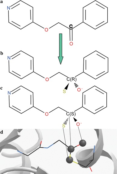
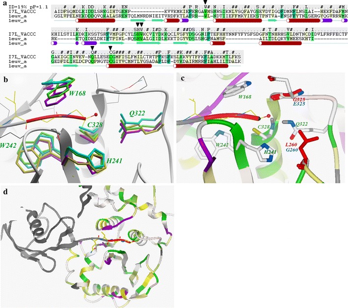
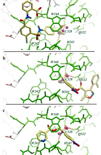
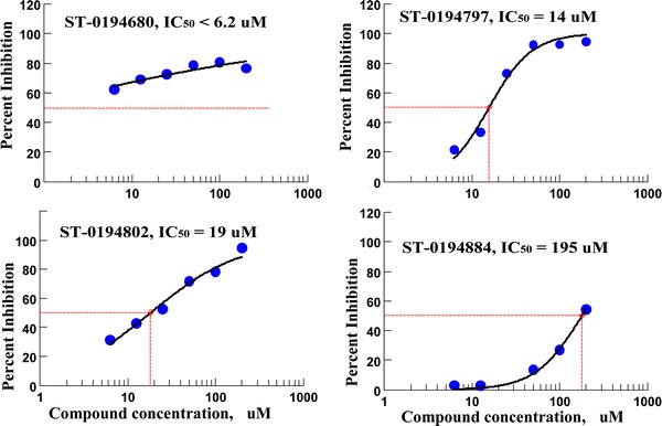
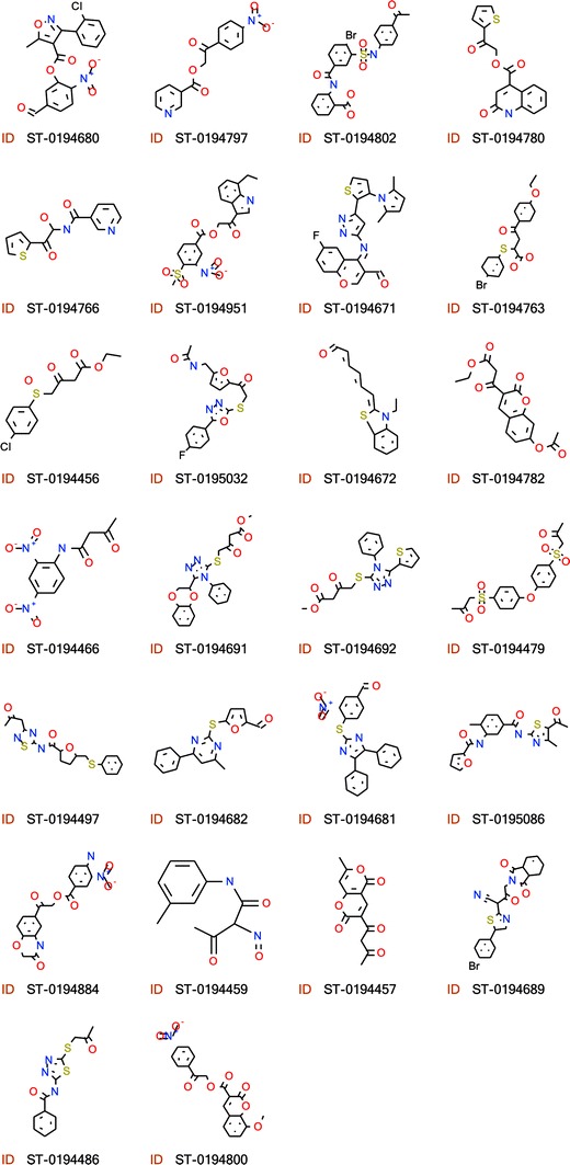
Essential for viral replication and highly conserved among poxviridae, the vaccinia virus I7L ubiquitin-like proteinase (ULP) is an attractive target for development of smallpox antiviral drugs. At the same time, the I7L proteinase exemplifies several interesting challenges from the rational drug design perspective. In the absence of a published I7L X-ray structure, we have built a detailed 3D model of the I7L ligand binding site (S2-S2' pocket) based on exceptionally high structural conservation of this site in proteases of the ULP family. The accuracy and limitations of this model were assessed through comparative analysis of available X-ray structures of ULPs, as well as energy based conformational modeling. The 3D model of the I7L ligand binding site was used to perform covalent docking and VLS of a comprehensive library of about 230,000 available ketone and aldehyde compounds. Out of 456 predicted ligands, 97 inhibitors of I7L proteinase activity were confirmed in biochemical assays ( approximately 20% overall hit rate). These experimental results both validate our I7L ligand binding model and provide initial leads for rational optimization of poxvirus I7L proteinase inhibitors. Thus, fragments predicted to bind in the prime portion of the active site can be combined with fragments on non-prime side to yield compounds with improved activity and specificity.
Figures
Similar articles
-
Molecular dissection of the vaccinia virus I7L core protein proteinase.J Virol. 2003 Oct;77(20):11279-83. doi: 10.1128/jvi.77.20.11279-11283.2003. J Virol. 2003. PMID: 14512576 Free PMC article.
-
Dynamic Cap-Mediated Substrate Access and Potent Inhibitor Design of Monkeypox Virus I7L Protease.Adv Sci (Weinh). 2025 Jul;12(26):e2501625. doi: 10.1002/advs.202501625. Epub 2025 Apr 7. Adv Sci (Weinh). 2025. PMID: 40193298 Free PMC article.
-
The vaccinia virus I7L gene product is the core protein proteinase.J Virol. 2002 Sep;76(17):8973-6. doi: 10.1128/jvi.76.17.8973-8976.2002. J Virol. 2002. PMID: 12163618 Free PMC article.
-
The SARS-coronavirus papain-like protease: structure, function and inhibition by designed antiviral compounds.Antiviral Res. 2015 Mar;115:21-38. doi: 10.1016/j.antiviral.2014.12.015. Epub 2014 Dec 29. Antiviral Res. 2015. PMID: 25554382 Free PMC article. Review.
-
Papain-like lysosomal cysteine proteases and their inhibitors: drug discovery targets?Biochem Soc Symp. 2003;(70):15-30. doi: 10.1042/bss0700015. Biochem Soc Symp. 2003. PMID: 14587279 Review.
Cited by
-
Assignment of pterin deaminase activity to an enzyme of unknown function guided by homology modeling and docking.J Am Chem Soc. 2013 Jan 16;135(2):795-803. doi: 10.1021/ja309680b. Epub 2013 Jan 2. J Am Chem Soc. 2013. PMID: 23256477 Free PMC article.
-
Virtual ligand screening against comparative protein structure models.Methods Mol Biol. 2012;819:105-26. doi: 10.1007/978-1-61779-465-0_8. Methods Mol Biol. 2012. PMID: 22183533 Free PMC article.
-
Drug design for ever, from hype to hope.J Comput Aided Mol Des. 2012 Jan;26(1):137-50. doi: 10.1007/s10822-011-9519-9. Epub 2012 Jan 18. J Comput Aided Mol Des. 2012. PMID: 22252446 Free PMC article.
-
Molecular docking screens using comparative models of proteins.J Chem Inf Model. 2009 Nov;49(11):2512-27. doi: 10.1021/ci9003706. J Chem Inf Model. 2009. PMID: 19845314 Free PMC article.
-
In silico identification of potential phytochemical inhibitors for mpox virus: molecular docking, MD simulation, and ADMET studies.Mol Divers. 2024 Dec;28(6):4067-4086. doi: 10.1007/s11030-023-10797-2. Epub 2024 Mar 22. Mol Divers. 2024. PMID: 38519803
References
Publication types
MeSH terms
Substances
Grants and funding
LinkOut - more resources
Full Text Sources
