. 2007 Oct 25:7:200.
doi: 10.1186/1471-2148-7-200.

Visual pigments in a living fossil, the Australian lungfish Neoceratodus forsteri

Affiliations

Visual pigments in a living fossil, the Australian lungfish Neoceratodus forsteri

Helena J Bailes et al. BMC Evol Biol. .

Abstract

Background: One of the greatest challenges facing the early land vertebrates was the need to effectively interpret a terrestrial environment. Interpretation was based on ocular adaptations evolved for an aquatic environment millions of years earlier. The Australian lungfish Neoceratodus forsteri is thought to be the closest living relative to the first terrestrial vertebrate, and yet nothing is known about the visual pigments present in lungfish or the early tetrapods.

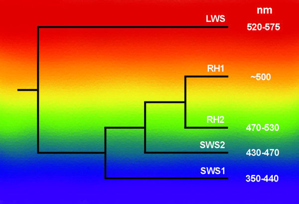
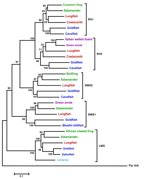
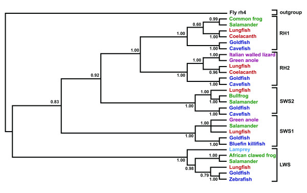
Results: Here we identify and characterise five visual pigments (rh1, rh2, lws, sws1 and sws2) expressed in the retina of N. forsteri. Phylogenetic analysis of the molecular evolution of lungfish and other vertebrate visual pigment genes indicates a closer relationship between lungfish and amphibian pigments than to pigments in teleost fishes. However, the relationship between lungfish, the coelacanth and tetrapods could not be absolutely determined from opsin phylogeny, supporting an unresolved trichotomy between the three groups.

Conclusion: The presence of four cone pigments in Australian lungfish suggests that the earliest tetrapods would have had a colorful view of their terrestrial environment.


Figures

Figure 1
Figure 1
The phylogenetic relationships between the vertebrate visual opsin lineages. A series of four duplication events produced the lws, sws1, sws2 and then rh1 and rh2 genes. The position of each branch on the spectrum portrays the approximate spectral sensitivity of each opsin group. Maximum absorbance value ranges (nm) are based on pigments reconstructed with 11-cis retinal. Values are taken from [45], figure adapted from [3].
Figure 2
Figure 2
Multiple transcripts of rh1 occur in the Australian lungfish retina. A) 3' RACE PCR products from lungfish rh1 cDNA. Arrows point to two potential transcripts differing in size in lane 2 (a third, faint band is also visible above these two but was not successfully sequenced), while lane 3 contained no template cDNA. The vertical grey line indicates where an irrelevant region of gel was removed using Photoshop 6.0 (Adobe). B) The 3' UTR of lungfish rh1. Two sequenced polyA signals are present (bold and italicised), both of which can induce polyadenylation producing two differently sized Rh1 transcripts.
Figure 3
Figure 3
A phylogenetic tree of the five photoreceptor opsins of Neoceratodus forsteri and selected full-length nucleotide coding sequences of related species. The tree was constructed using the Neighbour-joining method with 1000 bootstrap replications [39]. Sarcopterygian fish (coelacanth and lungfish) are in red, agnathan fish (lamprey) are in light blue, teleost fish are in dark blue, amphibians are in green and reptiles are in purple. Genbank accession numbers are listed in Table 1. Bootstrap confidence values are at the base of each node. The rh4 opsin of Drosophila melanogaster (Table 1) was used as an outgroup. Scale bar indicates nucleotide substitutions per site.
Figure 4
Figure 4
A clade credibility tree showing the relationships between lungfish and other selected vertebrate opsins. The tree was generated using Bayesian inference via a Metroplis-coupled Markov chain Monte Carlo simulation. Sarcopterygian fish (coelacanth and lungfish) are in red, teleost fish are in blue, amphibians are in green and reptiles are in maroon. Genbank accession numbers are listed in Table 1. Posterior probability values are at the base of each node. The rh4 opsin of Drosophila melanogaster is used as an outgroup (Table 1). The probability of most relationships within the tree is 1.00 after 300,000 generations, while lower posterior probability values are found within the lws and rh1 groups.

Similar articles

Cited by

References

    1. Yokoyama S, Yokoyama R. Adaptive evolution of photoreceptors and visual pigments in vertebrates. Annu Rev Ecol Syst. 1996;27:543–567. doi: 10.1146/annurev.ecolsys.27.1.543. - DOI
    1. Spady TC, Seehausen O, Loew ER, Jordan RC, Kocher TD, Carleton KL. Adaptive molecular evolution in the opsin genes of rapidly speciating Cichlid species. Mol Biol Evol. 2005;22:1412–1422. doi: 10.1093/molbev/msi137. - DOI - PubMed
    1. Trezise AEO, Collin SP. Opsins: Evolution in waiting. Curr Biol. 2005;15:R794–R796. doi: 10.1016/j.cub.2005.09.025. - DOI - PubMed
    1. Nathans J, Thomas D, Hogness DS. Molecular genetics of human color vision: the genes encoding blue, green, and red pigments. Science. 1986;232:193–202. doi: 10.1126/science.2937147. - DOI - PubMed
    1. Johnson RL, Grant KB, Zankel TC, Boehm MF, Merbs SL, Nathans J, Nakanishi K. Cloning and Expression of Goldfish Opsin Sequences. Biochemistry. 1993;32:208–214. doi: 10.1021/bi00052a027. - DOI - PubMed

Publication types

LinkOut - more resources