. 2007 Oct 25:7:201.
doi: 10.1186/1471-2148-7-201.

Dicyema Pax6 and Zic: tool-kit genes in a highly simplified bilaterian

Affiliations

Dicyema Pax6 and Zic: tool-kit genes in a highly simplified bilaterian

Jun Aruga et al. BMC Evol Biol. .

Abstract

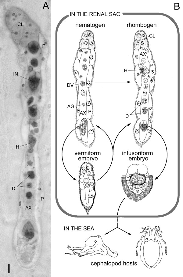
Background: Dicyemid mesozoans (Phylum Dicyemida) are simple (8-40-cell) cephalopod endoparasites. They have neither body cavities nor differentiated organs, such as nervous and gastrointestinal systems. Whether dicyemids are intermediate between Protozoa and Metazoa (as represented by their "Mesozoa" classification) or degenerate species of more complex metazoans is controversial. Recent molecular phylogenetic studies suggested that they are simplified bilaterians belonging to the Lophotrochozoa. We cloned two genes developmentally critical in bilaterian animals (Pax6 and Zic), together with housekeeping genes (actin, fructose-bisphosphate aldolase, and ATP synthase beta subunit) from a dicyemid to reveal whether their molecular phylogeny supported the "simplification" hypothesis, and to clarify evolutionary changes in dicyemid gene structure and expression profiles.

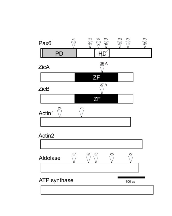
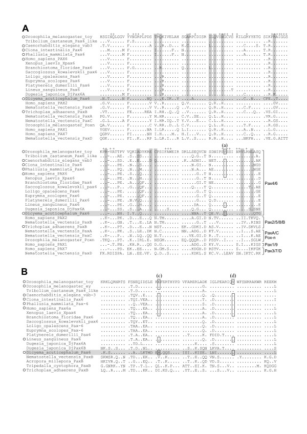
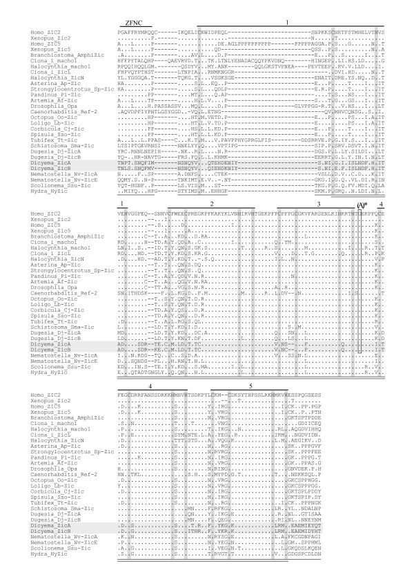
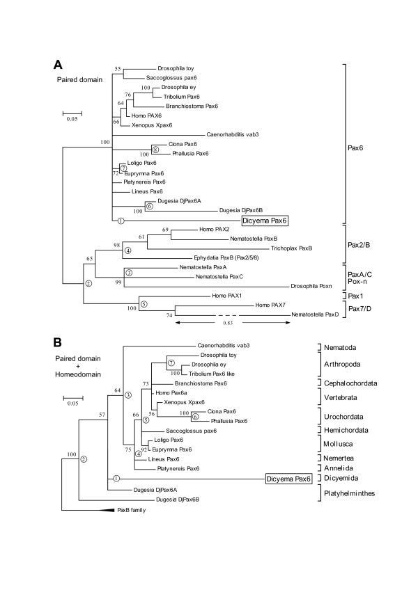
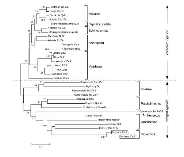
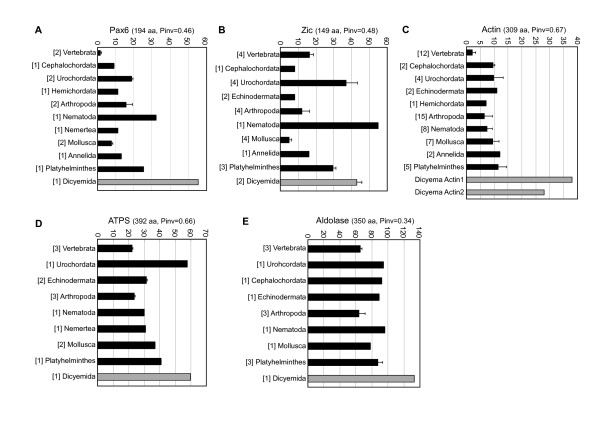
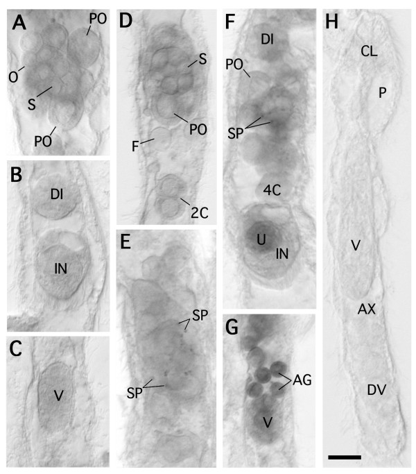
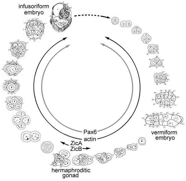
Results: Genomic/cDNA sequence analysis showed that 1) the Pax6 molecular phylogeny and Zic intron positions supported the idea of dicyemids as reduced bilaterians; 2) the aa sequences deduced from the five genes were highly divergent; and 3) Dicyema genes contained very short introns of uniform length. In situ hybridization analyses revealed that Zic genes were expressed in hermaphroditic gonads, and Pax6 was expressed weakly throughout the developmental stages of the 2 types of embryo and in the hermaphroditic gonads.

Conclusion: The accelerated evolutionary rates and very short and uniform intron may represent a part of Dicyema genomic features. The presence and expression of the two tool-kit genes (Pax6 and Zic) in Dicyema suggests that they can be very versatile genes even required for the highly reduced bilaterian like Dicyema. Dicyemids may be useful models of evolutionary body plan simplification.


Figures

Figure 1
Figure 1
Dicyema acuticephalum. (A) Light micrograph of a rhombogen. Note that embryos develop in the axial cell. Scale bar, 10 μm. (B) Life cycle of the dicyemid (after Furuya and Tsuneki [1]). The dotted line indicates an unknown process. The vermiform stage includes the nematogen, rhombogen, and vermiform embryo. The development of infusorigens, gametogenesis around the infusorigen, and development of the 2 types of embryo all proceed within the axial cell.AG, agamete; AX, axial cell; CL, calotte; D, developing infusoriform embryo; DV, developing vermiform embryo;H, hermaphroditic gonad (infusorigen); IN, infusoriform embryo; P, peripheral cell.
Figure 2
Figure 2
Dicyemid genes cloned in this study. The entire protein-encoding (open box) domains and the functional domains (ZF, zinc finger domain, black; PD, paired box, gray; HD, homeodomain, hatched) are drawn proportionally. Positions of introns are indicated by open triangles. The number above each open triangle indicates the size (in nucleotides) of the intron at that position. actin2 and ATP synthase lack introns. Scale bar, 100 aa.
Figure 3
Figure 3
Multiple alignment of Pax6 aa sequences. The sequences corresponding to Pax6 PD (A) and HD (B) were aligned. Dots indicate aa residues identical to those of the top line (Drosophila_toy).Dashes indicate artificial spaces inserted by the alignment program. + indicates the residue that interact with DNA [14]. Horizontal shaded lines indicate the Dicyema sequence. Vertical shaded lines indicate aa residues strongly conserved in Pax6 subfamily, but not in the others.Open boxes represent the positions of introns. The phases of introns are (a), 0; (c), 1; (d), 0. Open circles indicate animals in which intron positions were examined.
Figure 4
Figure 4
Multiple alignment of aa sequences corresponding to Zic ZF and flanking regions. Dots represent aa residues identical to those in the top lines (Homo_ZIC2). Dashes indicate artificial spaces inserted by the alignment program. Horizontal shaded lines indicate the Dicyema Zic sequences. Vertical shaded lines indicate the positions of cysteine and histidine residues in the C2H2 motif. Labels above the top lines indicate subregions (ZFNC, Zinc finger N-terminal conserved region; 1–5, C2H2 ZF motif 1–5). Open box labeled with (A)* indicates the position of the bilaterian-specific intron (A-intron) [15], where the exon-intron boundaries for the Halocynthia, Octopus, and Dugesia sequences have not yet been clarified. Cnidarian Zic genes (Nematostella, Scolionema, and Hydra) lack the intron in this position. Bottom parallel lines indicate the region subjected to the aa substitution rate analysis. Bottom underlines and parallel lines indicate the region used for the NJ tree analysis.
Figure 5
Figure 5
BI tree based on metazoan Pax6-related aa sequences. BI trees of Pax6 were drawn by MrBayes [44] by using PD sequences (A) and concatenated PD+HD sequences (PD+HD). PD tree was unrooted while PD+HD tree was rooted with PaxB family of which subtree is indicated in closed triangle. Numbers next to interior branches indicate posterior probability. Circled numbers indicate the Dicyema-branch-regrafted position in the alternative tree analyses (Tables 1 and 2). Scale bar represents evolutionary distance in substitutions/aa residue. Classification of Pax genes according to a previous study [20] is indicated in the right side of (A).
Figure 6
Figure 6
NJ tree based on metazoan Zic-related aa sequences. NJ tree of Zic was drawn by MEGA3 [43] by using Zic ZF core sequences (double underlined region in Fig. 4). The evolutionary distances were determined using the JTT matrix [46]. The internal labels indicate the percentage value of bootstrap test (1000 replicates). Scale bar represents evolutionary distance in substitutions/aa residue. Classification of Zic genes according to a previous study [15] is indicated by broken line and arrows.
Figure 7
Figure 7
Amino acid substitution rates in relation to bilaterian ancestor sequences. In each graph, the x-axes indicate mean numbers of substituted aa in comparison to ancestor sequences in each taxa, with standard deviations (error bars). A, Pax6 PD+HD; B, Zic ZF; C, actin; D, ATPS; E, Aldolase. Bracketed numbers left of the taxa names indicate the numbers of sequences subjected to the analysis. aa, length of aa sequences subjected to the analysis; Pinv, proportion of invariant aa among all tested sequences. The animal species are listed with their sequence accession numbers [see Additional file 2].
Figure 8
Figure 8
In situ hybridization of dicyemid ZicA, ZicB, Pax6, and actin1. (A-C) ZicA; (D) ZicB; (E) Pax6; (F, G) actin1; (H) in situ hybridization, without RNA probe as a negative control. (A) infusorigen; (B) later stage of infusoriform embryo and formed infusoriform embryo; (C) later stage of vermiform embryo; (D) infusorigen and 2-cell-stage embryo; (E), infusorigen; (F), infusorigen and early stages of developing infusoriform embryos, and formed infusoriform embryo; (G), agametes and later stage of vermiform embryo; (H), whole body of young individual. Scale bar, 10 μm. AG, agamete; AX, axial cell; CL, calotte; DI, developing infusoriform embryo; DV, developing vermiform embryo; F, fertilized egg; IN, infusoriform embryo; O, oogonium; PO, primary oocyte; P, peripheral cell; S, spermatogonium; SP, sperm; U, urn cell; V, vermiform embryo; 2C, 2-cell-stage embryo; 4C, 4-cell-stage embryo.
Figure 9
Figure 9
Expression patterns of ZicA, ZicB, Pax6, and actin in developmental processes of D. acuticephalum. The development of the hermaphroditic gonad, vermiform embryo, and infusoriform embryo is based on Furuya et al. [50, 51, 52].

Similar articles

Cited by

References

    1. Furuya H, Tsuneki K. Biology of dicyemid mesozoans. Zoolog Sci. 2003;20:519–532. doi: 10.2108/zsj.20.519. - DOI - PubMed
    1. Furuya H, Hochberg FG, Tsuneki K. Developmental patterns and cell lineages of vermiform embryos in dicyemid mesozoans. Biol Bull. 2001;201:405–416. doi: 10.2307/1543618. - DOI - PubMed
    1. Katayama T, Wada H, Furuya H, Satoh N, Yamamoto M. Phylogenetic position of the dicyemid mesozoa inferred from 18S rDNA sequences. Biol Bull. 1995;189:81–90. doi: 10.2307/1542458. - DOI - PubMed
    1. Kobayashi M, Furuya H, Holland PW. Dicyemids are higher animals. Nature. 1999;401:762. doi: 10.1038/43273. - DOI - PubMed
    1. Gehring WJ. The genetic control of eye development and its implications for the evolution of the various eye-types. Int J Dev Biol. 2002;46:65–73. - PubMed

Publication types

MeSH terms

LinkOut - more resources