. 2007 Oct 25:8:96.
doi: 10.1186/1471-2199-8-96.

Regulation of poly(ADP-ribose) polymerase-1 (PARP-1) gene expression through the post-translational modification of Sp1: a nuclear target protein of PARP-1

Affiliations

Regulation of poly(ADP-ribose) polymerase-1 (PARP-1) gene expression through the post-translational modification of Sp1: a nuclear target protein of PARP-1

Karine Zaniolo et al. BMC Mol Biol. .

Abstract

Background: Poly(ADP-ribose) polymerase-1 (PARP-1) is a nuclear enzyme that plays critical functions in many biological processes, including DNA repair and gene transcription. The main function of PARP-1 is to catalyze the transfer of ADP-ribose units from nicotinamide adenine dinucleotide (NAD+) to a large array of acceptor proteins, which comprises histones, transcription factors, as well as PARP-1 itself. We have previously demonstrated that transcription of the PARP-1 gene essentially rely on the opposite regulatory actions of two distinct transcription factors, Sp1 and NFI. In the present study, we examined whether suppression of PARP-1 expression in embryonic fibroblasts derived from PARP-1 knockout mice (PARP-1-/-) might alter the expression and/or DNA binding properties of Sp1 and NFI. We also explored the possibility that Sp1 or NFI (or both) may represent target proteins of PARP-1 activity.

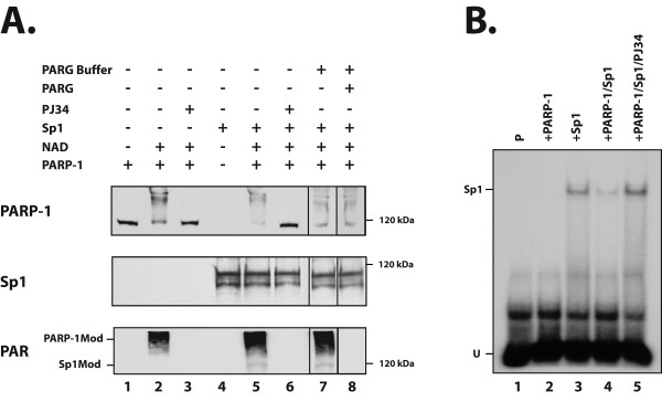
Results: Expression of both Sp1 and NFI was found to be considerably reduced in PARP-1-/- cells. Co-immunoprecipitation assays revealed that PARP-1 physically interacts with Sp1 in a DNA-independent manner, but neither with Sp3 nor NFI, in PARP-1+/+ cells. In addition, in vitro PARP assays indicated that PARP-1 could catalyze the addition of polymer of ADP-ribose to Sp1, which also translated into a reduction of Sp1 binding to its consensus DNA target site. Transfection of the PARP-1 promoter into both PARP-1+/+ and PARP-1-/- cells revealed that the lack of PARP-1 expression in PARP-1-/- cells also results in a strong increase in PARP-1 promoter activity. This influence of PARP-1 was found to rely on the presence of the Sp1 sites present on the basal PARP-1 promoter as their mutation entirely abolished the increased promoter activity observed in PARP-1-/- cells. Subjecting PARP-1+/+ cells to an oxidative challenge with hydrogen peroxide to increase PARP-1 activity translated into a dramatic reduction in the DNA binding properties of Sp1. However, its suppression by the inhibitor PJ34 improved DNA binding of Sp1 and led to a dramatic increase in PARP-1 promoter function.

Conclusion: Our results therefore recognized Sp1 as a target protein of PARP-1 activity, the addition of polymer of ADP-ribose to this transcription factor restricting its positive regulatory influence on gene transcription.


Figures

Figure 1
Figure 1
Expression of PARP-1, Sp1, Sp3 and NFI in PARP-1+/+ and PARP-1-/- cells. Crude nuclear extracts (10 μg) from both PARP-1+/+ and PARP-1-/- cells were examined in Western blot using antibodies directed against PARP-1, Sp1, Sp3 and NFI. The position of the 120 kDa and 60 kDa proteins used as molecular mass markers is indicated. The asterisk indicates the position of the typical NFI complex whereas the arrowhead designates NFI complexes with a reduced electrophoretic mobility that predominated in the extract from PARP-1-/- cells. Data of one from three similar experiments are presented.
Figure 2
Figure 2
DNA binding properties of Sp1/Sp3 and NFI in PARP-1+/+ and PARP-1-/- cells. (A) EMSA analysis of Sp1/Sp3 and NFI. Crude nuclear proteins (5 μg) from both PARP-1+/+ and PARP-1-/- cells were incubated with a 5' end-labeled probe bearing the high affinity binding site for either Sp1 (left) or NFI (right). Formation of DNA/protein complexes was then monitored by EMSA on an 8% (Sp1) and 10% (NFI) native polyacrylamide gel and their position revealed through autoradiography. The position of both the Sp1/Sp3 and NFI DNA-protein complexes are shown, as well as that of the free probe (U). P: labeled probe alone. (B) Sp1 competition experiment in EMSA. The Sp1 labeled probe used in panel A was incubated with nuclear proteins (5 μg) from both PARP-1+/+ and PARP-1-/- cells in the presence of either no (-) or 100- and 500-fold molar excesses of unlabeled competitor oligonucleotides (either Sp1 or NFI). Formation of DNA/protein complexes was then monitored by EMSA on an 8% native gel. (C) NFI competition experiment in EMSA. Same as in panel B except that the NFI double-stranded oligonucleotide was 5'-end labeled and used as probe for the assay. (D) Supershift experiment in EMSA. Crude nuclear proteins from both PARP-1+/+ and PARP-1-/- cells were incubated with the either the Sp1 (5 μg proteins were used) or NFI (10 μg proteins were used) labeled probe in the presence of either no (-), or 2 μl of a polyclonal antibody directed against Sp1 (Sp1Ab) or Sp3 (Sp3Ab) and added either individually or in combination (Sp1+Sp3Ab) (left), or with a polyclonal antibody directed against NFI (right). Formation of both the Sp1/Sp3 and NFI complexes, as well as their corresponding supershifted complexes (SSC) is indicated. P: labeled probe alone; U: unbound fraction of the labeled probe.
Figure 3
Figure 3
DNA binding and expression of the transcription factors AP-1, E2F1 and STAT-1 in PARP-1+/+ and PARP-1-/- cells. (A) EMSA analysis. Crude nuclear proteins (5 μg) from both PARP-1+/+ and PARP-1-/- cells were incubated with a 5' end-labeled probe bearing the high affinity binding site for AP-1, E2F1 and STAT-1. Formation of DNA/protein complexes was then monitored by EMSA on an 8% gel as detailed in Figure 2. The position of the AP-1, E2F1 and STAT-1 DNA-protein complexes is shown, as well as that of the free probe (U). P: labeled probe alone. (B) Coomassie blue staining of the protein samples used for conducting both the EMSA (panel A) and the Western blot experiment (panel C). One protein band present in both extract was randomly selected and its intensity determined by densitometric analysis in order to precisely calibrate the protein concentration used for the assays. (C) Nuclear extracts (10 μg) from both PARP-1+/+ and PARP-1-/- cells were examined in Western blot as in Figure 1 using antibodies directed against E2F1, STAT-1 and the AP-1 subunit c-jun. The position of the nearest molecular mass markers is indicated (60 kDa and 85 kDa).
Figure 4
Figure 4
Co-immunoprecipitation of Sp1 and PARP-1 in protein extracts from PARP-1+/+ and PARP-1-/- cells. (A) Immunoprecipitation of the Sp1-protein complexes in PARP-1+/+ and PARP-1-/- nuclear extracts. Crude nuclear proteins (300 μg) from both PARP-1+/+ and PARP-1-/- cells were incubated with the Sp1 Ab (sc-59) and the Sp1-protein complexes recovered by the addition of protein-A-Sepharose. The resulting immunoprecipitated proteins were then SDS-gel fractionated before being membrane-transferred and Western blotted with antibodies against Sp1, PARP-1 (C-2-10) and PAR (LP-9610). Ctl-: protein A-Sepharose added to crude nuclear proteins in the absence of Sp1 Ab and used as a negative control. IgG-Ab: normal rabbit IgG incubated with nuclear proteins prior to addition of protein A-Sepharose as a negative control. (B) Immunoprecipitation of the PARP-1-protein complexes in PARP-1+/+ and PARP-1-/- nuclear extracts. Same as in panel A except that the immunoprecipitation was conducted using the PARP-1 F-123 Ab. The blotted, PARP-1-immunoprecipitated proteins were then analyzed with the PARP-1 (422), Sp1 (sc-59), Sp3 (sc-644), and PAR (LP-9610) antibodies. Negative controls (Ctl- and IgG-Ab) are as in panel A. TE: total cell extract that has not been immunoprecipitated with the PARP-1 Ab.
Figure 5
Figure 5
Influence of PARP-1 activity on the co-immunoprecipitation of PARP-1 by Sp1. Nuclear proteins (300 μg) from PARP-1+/+ cells grown either alone (control) or in the presence of hydrogen peroxide (H2O2), PJ34 PARP-1 inhibitor, or ethidium bromide were incubated with the Sp1 Ab (sc-59) and the Sp1-protein complexes recovered by the addition of protein-A-Sepharose. The resulting immunoprecipitated proteins were then gel fractionated as in Figure 4 and Western blotted with antibodies against Sp1 or PARP-1 (C-2-10). TE: total cell extract that has not been immunoprecipitated with the Sp1 Ab. Ctl-: protein A-Sepharose added to crude nuclear proteins in the absence of Sp1 Ab and used as a negative control. IgG-Ab: normal rabbit IgG incubated with nuclear proteins prior to addition of protein A-Sepharose as a negative control.
Figure 6
Figure 6
PARP-1-dependent poly(ADP-ribosyl)ation of Sp1 in vitro. (A) Recombinant Sp1 protein was incubated in reaction buffer either alone (lane 4) or with purified bovine PARP-1 (1 unit) in the presence of 200 μM NAD+ (lane 5). The reaction mixture was subjected to Western blot analysis with the PARP-1 (C-2-10), Sp1 (sc-59) and PAR (LP-9610) antibodies. When indicated, the PARP inhibitor PJ34 was added to the reaction mixture with purified PARP-1 alone (lane 3) or in the presence of recombinant Sp1 (lane 6). When indicated, samples from the in vitro PARP assay were electrophoresed and electrotransfered onto nitrocellulose membranes. The PAR covalently linked onto the automodified PARP-1 and Sp1 proteins was then erased by incubation with PARG and the proteins analyzed by Western blotting with the same antibodies as detailed above (lane 8). Lane 1: PARP-1 alone; lane 2: PARP-1 incubated with NAD+; lane 3: same as in lane 2 plus PJ34; lane 7: same as in lane 5 but incubated in PARG buffer without addition of PARG-1. The position of modified PARP-1 (PARP-1Mod) and Sp1 (Sp1Mod) is indicated (left) along with the appropriate molecular mass marker (right). (B) Recombinant Sp1 was incubated in reaction buffer containing 200 μM NAD+ and nicked DNA either alone (+SP1; lane 3) or with purified bovine PARP-1 (1 unit) (+Sp1/PARP-1; lane 4). A sample (16 μl) from the reaction mixture was then incubated with the 5'-end labeled Sp1 oligonucleotide and formation of DNA-protein complexes monitored by EMSA as in Figure 2. As a control, the PARP-1 inhibitor PJ34 was added to the reaction mixture containing PARP-1/NAD+/Sp1 (+Sp1/PARP-1/PJ34; lane 5). Lane 1: labeled probe alone in reaction mix (P); Lane 2: labeled probe incubated in buffer D with PARP-1 but in the absence of NAD and Sp1 (+PARP-1). The position of both the Sp1 complex (Sp1) and the free probe (U) is indicated.
Figure 7
Figure 7
In vivo influence of PARP-1 activity on the expression and DNA binding of Sp1. (A) Nuclear proteins (5 μg) from PARP-1+/+ cells grown alone (-; lane 2) or in the presence of H2O2 (lane 3) or PJ34 (lane 4), added either individually or in combination (PJ34+ H2O2; lane 5), were incubated with the Sp1 labeled probe and formation of DNA/protein complexes monitored by EMSA on a 8% native polyacrylamide gel as in Figure 2. The position of both the Sp1 and Sp3 DNA-protein complexes are shown, as well as that of the free probe (U). P: labeled probe alone (lane 1). (B) The extracts used in panel A were SDS-gel fractionated before being membrane-transferred and Western blotted with antibodies against Sp1 (sc-59), PARP (C-2-10) and PAR (10-H). The position of the appropriate molecular mass markers (60-, 120-, and 190 kDa) is indicated. (C) Nuclear proteins (5 μg) from primary cultures of HSKs grown for various periods of time (4-, 24- and 72 h) either alone (-; lanes 1, 4 and 7), or in the presence of H2O2 (lanes 2, 5 and 8) or both H2O2 and PJ34 (PJ34+ H2O2; lanes 3, 6 and 9), were incubated with the Sp1 labeled probe and formation of DNA/protein complexes monitored by EMSA on a 8% native polyacrylamide gel as in panel A. (D) The extracts used in panel C were analyzed by Western blotting with antibodies against Sp1 (sc-59), PARP (C-2-10) and β-actin (CLT9001). Densitometric analyses of the band intensities was determined for both the Sp1 and PARP-1 proteins and normalized to that measured for β-actin. Values are shown below each corresponding track.
Figure 8
Figure 8
rPARP-1 promoter activity in PARP-1+/+ and PARP-1-/- cells. (A) The recombinant plasmids PCR3 and PCR3F2/F3/F4m were transfected into both PARP-1+/+ and PARP-1-/- cells grown with or without the PARP-1 inhibitor PJ34. CAT activities were measured and normalized to the amount of hGH secreted into the culture medium. Values are expressed as ((%CAT activity/100 μg proteins)/ng hGH). Asterisks (*) indicate CAT activities from cells exposed to PJ34 that are statistically different from those measured when cells are transfected with pCR3 in the absence of inhibitor whereas † corresponds to CAT activities in PARP-1-/- cells that are statistically different from those measured in PARP-1+/+ cells (P < 0.005; paired samples, t-test). S.D. is also provided. (B) The plasmids PCR3, -92α5CAT and α6–84 were transfected into both PARP-1+/+ and PARP-1-/- cells and CAT activity (expressed as the ratio of CAT activity from PARP-1-/- cells over that measured in PARP-1+/+ cells (considered as 100%)) measured and normalized as detailed above. Asterisks (*) correspond to CAT activities in PARP-1-/- cells that are statistically different from those measured in PARP-1+/+ cells (P < 0.005; paired samples, t-test).
Figure 9
Figure 9
Model of interplay between PARP-1, Sp1 and other transcription factors. (A) PARP-1 plays a suppressive function (indicated by 'T' bars) on the DNA binding properties of Sp1, and indirectly, on its expression as well, by the enzymatic addition of poly(ADP-ribose) units (PAR) to Sp1. PARP-1 may exert its effect by stimulating the transcriptional properties (indicated by arrows) of both AP-2 and E2F-1 by physically interacting with these transcription factors (and therefore, independently of addition of PAR), of which the latter was recognized as a component required to ensure proper transcription of the human Sp1 gene. (B) Once PARP-1 is stimulated by DNA damages, post-translational modification of both Sp1 and AP-2 is increased to the point that their DNA binding properties and thereby, their transcriptional capacity, is considerably decreased without significantly altering their level of expression. (C) However, in the absence of PARP-1, addition of PAR is abrogated and the transcriptional capacity of Sp1 becomes dramatically increased despite that its overall expression is considerably reduced primarily as a consequence of: i) a reduction in both the expression [112] and the positive transcriptional influence of E2F1 [85], a property that requires a physical interaction with PARP-1, and ii) a reduced transcriptional activity of AP-2, which also requires a physical association of this transcription factor with the middle region of PARP-1 [84]. TrC: transcriptional capacity of Sp1; Exp: level of Sp1 expression.

Similar articles

Cited by

References

    1. D'Amours D, Desnoyers S, D'Silva I, Poirier GG. Poly(ADP-ribosyl)ation reactions in the regulation of nuclear functions. Biochem J. 1999;342:249–268. doi: 10.1042/0264-6021:3420249. - DOI - PMC - PubMed
    1. Virag L, Szabo C. The therapeutic potential of poly(ADP-ribose) polymerase inhibitors. Pharmacol Rev. 2002;54:375–429. doi: 10.1124/pr.54.3.375. - DOI - PubMed
    1. Chatterjee S, Berger SJ, Berger NA. Poly(ADP-ribose) polymerase: a guardian of the genome that facilitates DNA repair by protecting against DNA recombination. Mol Cell Biochem. 1999;193:23–30. doi: 10.1023/A:1006983304079. - DOI - PubMed
    1. d'Adda di Fagagna F, Hande MP, Tong WM, Lansdorp PM, Wang ZQ, Jackson SP. Functions of poly(ADP-ribose) polymerase in controlling telomere length and chromosomal stability. Nat Genet. 1999;23:76–80. doi: 10.1038/12680. - DOI - PubMed
    1. de Murcia G, Menissier de Murcia J. Poly(ADP-ribose) polymerase: a molecular nick-sensor. Trends Biochem Sci. 1994;19:172–176. doi: 10.1016/0968-0004(94)90280-1. - DOI - PubMed

Publication types

MeSH terms

LinkOut - more resources