

Identification of novel ciliogenesis factors using a new in vivo model for mucociliary epithelial development
- PMID: 17961536
- PMCID: PMC2225594
- DOI: 10.1016/j.ydbio.2007.09.031
Identification of novel ciliogenesis factors using a new in vivo model for mucociliary epithelial development
Abstract
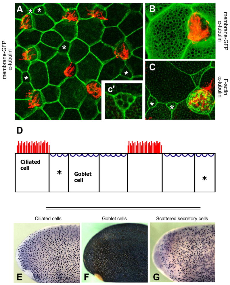
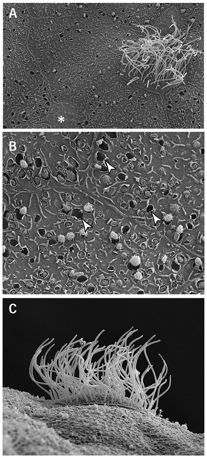
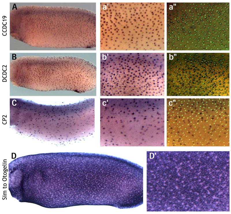
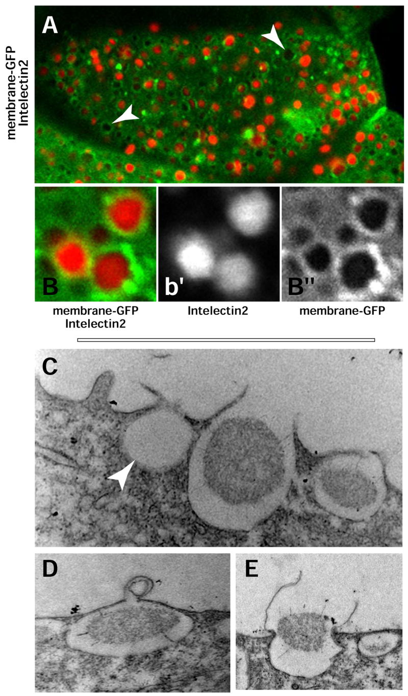
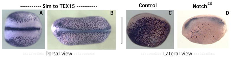
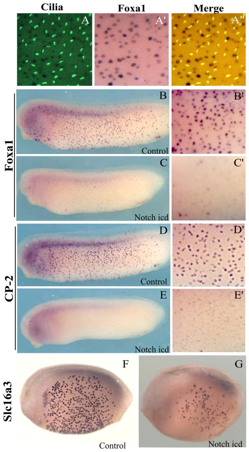
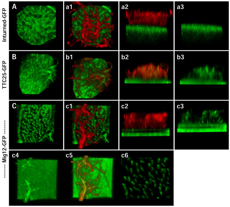
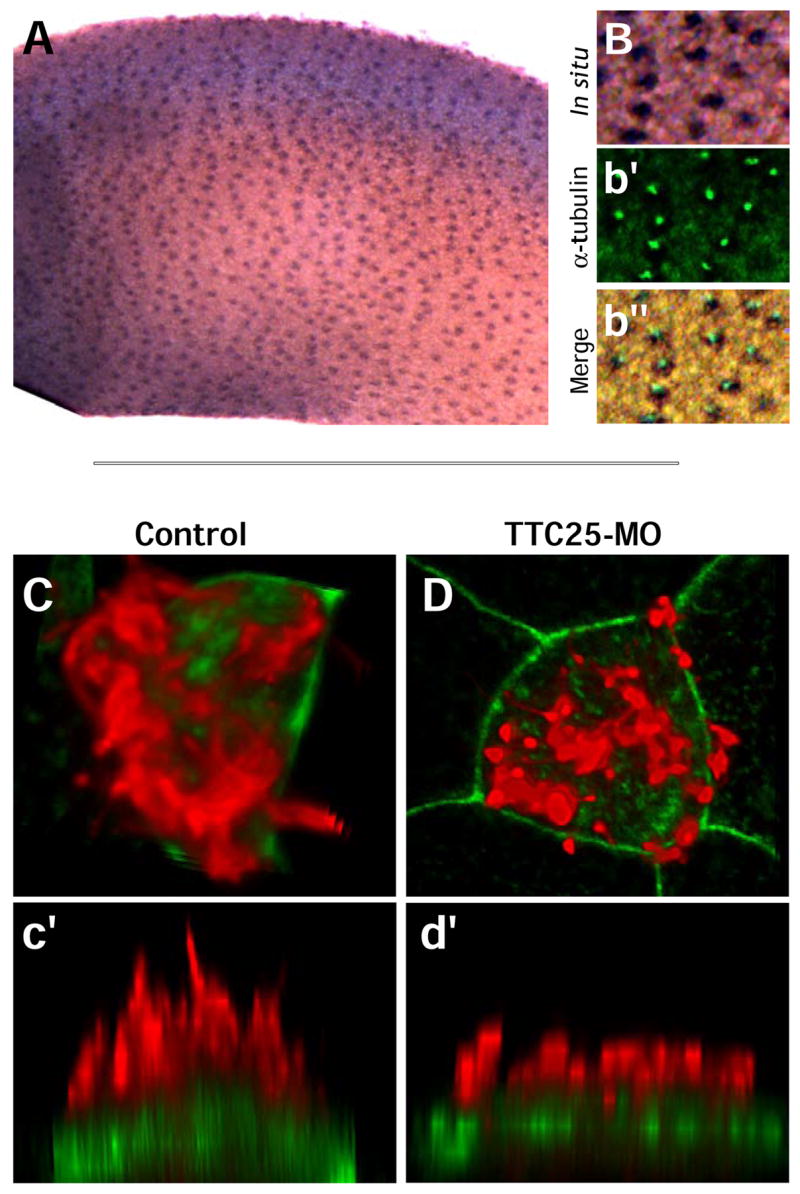
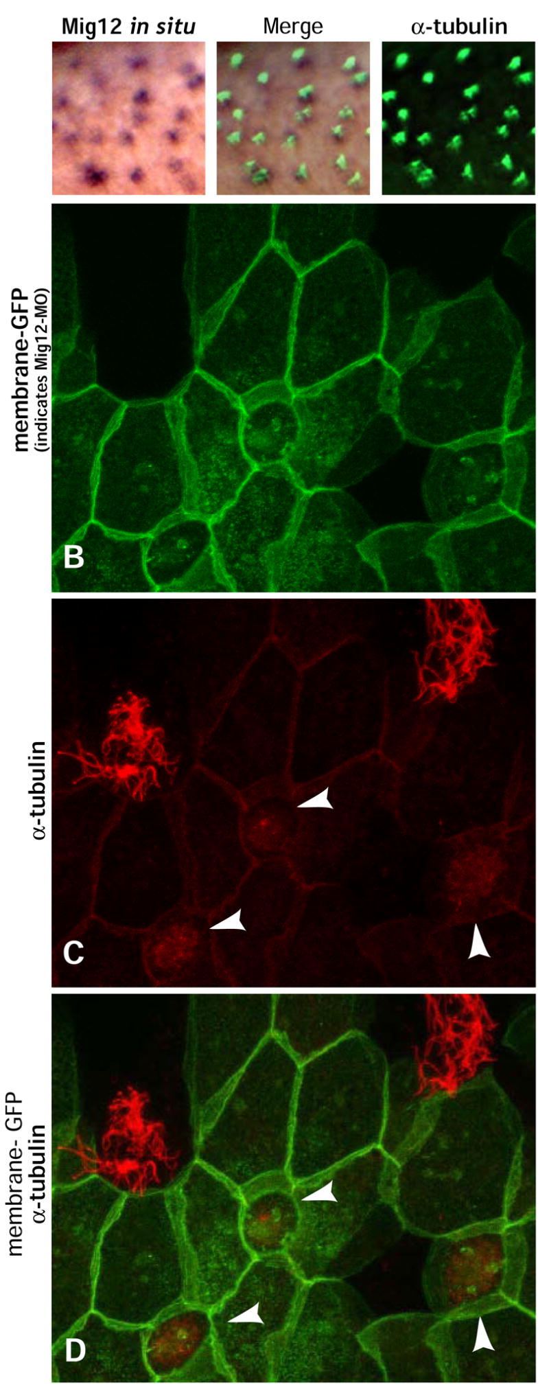
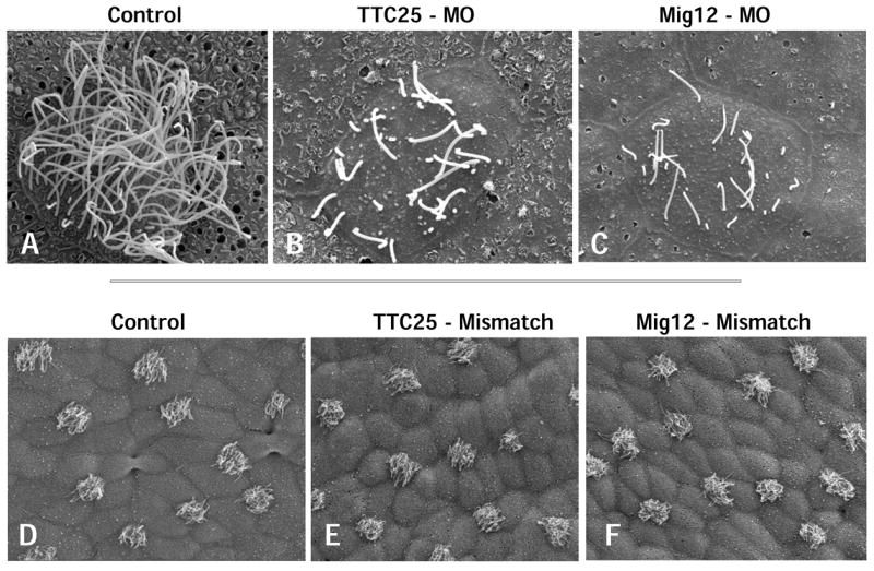
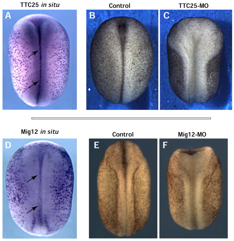
Mucociliary epithelia are essential for homeostasis of many organs and consist of mucus-secreting goblet cells and ciliated cells. Here, we present the ciliated epidermis of Xenopus embryos as a facile model system for in vivo molecular studies of mucociliary epithelial development. Using an in situ hybridization-based approach, we identified numerous genes expressed differentially in mucus-secreting cells or in ciliated cells. Focusing on genes expressed in ciliated cells, we have identified new candidate ciliogenesis factors, including several not present in the current ciliome. We find that TTC25-GFP is localized to the base of cilia and to ciliary axonemes, and disruption of TTC25 function disrupts ciliogenesis. Mig12-GFP localizes very strongly to the base of cilia and confocal imaging of this construct allows for simple visualization of the planar polarity of basal bodies that underlies polarized ciliary beating. Knockdown of Mig12 disrupts ciliogenesis. Finally, we show that ciliogenesis factors identified in the Xenopus epidermis are required in the midline to facilitate neural tube closure. These results provide further evidence of a requirement for cilia in neural tube morphogenesis and suggest that genes identified in the Xenopus epidermis play broad roles in ciliogenesis. The suites of genes identified here will provide a foundation for future studies, and may also contribute to our understanding of pathological changes in mucociliary epithelia that accompany diseases such as asthma.
Figures
Similar articles
-
RFX2 is broadly required for ciliogenesis during vertebrate development.Dev Biol. 2012 Mar 1;363(1):155-65. doi: 10.1016/j.ydbio.2011.12.029. Epub 2011 Dec 29. Dev Biol. 2012. PMID: 22227339 Free PMC article.
-
Embryonic frog epidermis: a model for the study of cell-cell interactions in the development of mucociliary disease.Dis Model Mech. 2011 Mar;4(2):179-92. doi: 10.1242/dmm.006494. Epub 2010 Dec 23. Dis Model Mech. 2011. PMID: 21183475 Free PMC article.
-
CFAP43 modulates ciliary beating in mouse and Xenopus.Dev Biol. 2020 Mar 15;459(2):109-125. doi: 10.1016/j.ydbio.2019.12.010. Epub 2019 Dec 27. Dev Biol. 2020. PMID: 31884020
-
Understanding ciliated epithelia: the power of Xenopus.Genesis. 2012 Mar;50(3):176-85. doi: 10.1002/dvg.20824. Epub 2011 Dec 27. Genesis. 2012. PMID: 22083727 Free PMC article. Review.
-
Xenopus epidermal and endodermal epithelia as models for mucociliary epithelial evolution, disease, and metaplasia.Genesis. 2021 Feb;59(1-2):e23406. doi: 10.1002/dvg.23406. Epub 2021 Jan 5. Genesis. 2021. PMID: 33400364 Review.
Cited by
-
Actin Depolymerizing Factor Destrin Regulates Cilia Development and Function during Vertebrate Embryogenesis.Dev Reprod. 2024 Sep;28(3):109-119. doi: 10.12717/DR.2024.28.3.109. Epub 2024 Sep 30. Dev Reprod. 2024. PMID: 39444639 Free PMC article.
-
ERK7 regulates ciliogenesis by phosphorylating the actin regulator CapZIP in cooperation with Dishevelled.Nat Commun. 2015 Mar 31;6:6666. doi: 10.1038/ncomms7666. Nat Commun. 2015. PMID: 25823377
-
RFX2 is broadly required for ciliogenesis during vertebrate development.Dev Biol. 2012 Mar 1;363(1):155-65. doi: 10.1016/j.ydbio.2011.12.029. Epub 2011 Dec 29. Dev Biol. 2012. PMID: 22227339 Free PMC article.
-
Xenopus embryonic epidermis as a mucociliary cellular ecosystem to assess the effect of sex hormones in a non-reproductive context.Front Zool. 2014 Feb 6;11(1):9. doi: 10.1186/1742-9994-11-9. Front Zool. 2014. PMID: 24502321 Free PMC article.
-
Embryonic frog epidermis: a model for the study of cell-cell interactions in the development of mucociliary disease.Dis Model Mech. 2011 Mar;4(2):179-92. doi: 10.1242/dmm.006494. Epub 2010 Dec 23. Dis Model Mech. 2011. PMID: 21183475 Free PMC article.
References
-
- Altmann CR, Bell E, Sczyrba A, Pun J, Bekiranov S, Gaasterland T, Brivanlou AH. Microarray-based analysis of early development in Xenopus laevis. Dev Biol. 2001;236:64–75. - PubMed
-
- Ansley SJ, Badano JL, Blacque OE, Hill J, Hoskins BE, Leitch CC, Kim JC, Ross AJ, Eichers ER, Teslovich TM, Mah AK, Johnsen RC, Cavender JC, Lewis RA, Leroux MR, Beales PL, Katsanis N. Basal body dysfunction is a likely cause of pleiotropic Bardet-Biedl syndrome. Nature. 2003;425:628–33. - PubMed
-
- Assheton R. Notes on the ciliation of the ectoderm of the amphibian embryo. Q J Microsc Sci. 1896;38:465–484.
Publication types
MeSH terms
Substances
Grants and funding
LinkOut - more resources
Full Text Sources
Other Literature Sources
