

Development and preclinical evaluation of an alphavirus replicon vaccine for influenza
- PMID: 17961878
- PMCID: PMC2706696
- DOI: 10.1016/j.vaccine.2007.09.038
Development and preclinical evaluation of an alphavirus replicon vaccine for influenza
Abstract
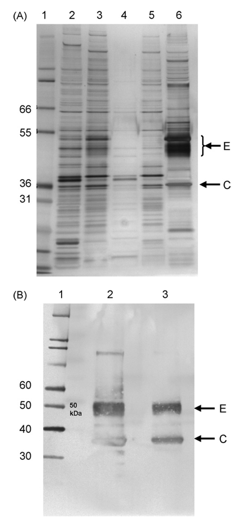
We used a propagation-defective, single-cycle, alphavirus replicon vector system to produce virus-like replicon particles (VRP) expressing the hemagglutinin (HA) and neuraminidase (NA) proteins from influenza A/Wyoming/03/2003 (H3N2). Efficient production methods were scaled to produce pilot lots of HA VRP and NA VRP and clinical lots of HA VRP. HA VRP-induced high-titered antibody responses in mice, rabbits and rhesus macaques, as measured by ELISA or hemagglutination inhibition (HI) assays, and robust cellular immune responses in mice and rhesus macaques, as measured by IFN-gamma ELISPOT. NA VRP also induced cellular immune responses in mice. A toxicology study with HA VRP and NA VRP in rabbits showed no adverse effects in any parameter. These studies support clinical testing of alphavirus replicon vaccines for influenza.
Figures


Similar articles
-
Development and preclinical evaluation of an alphavirus replicon particle vaccine for cytomegalovirus.Vaccine. 2007 Oct 16;25(42):7441-9. doi: 10.1016/j.vaccine.2007.08.016. Epub 2007 Aug 30. Vaccine. 2007. PMID: 17870214 Free PMC article.
-
Alphavirus replicon particle vaccines developed for use in humans induce high levels of antibodies to influenza virus hemagglutinin in swine: proof of concept.Vaccine. 2010 Jan 8;28(3):594-6. doi: 10.1016/j.vaccine.2009.10.015. Epub 2009 Oct 22. Vaccine. 2010. PMID: 19853679
-
Replicon-helper systems from attenuated Venezuelan equine encephalitis virus: expression of heterologous genes in vitro and immunization against heterologous pathogens in vivo.Virology. 1997 Dec 22;239(2):389-401. doi: 10.1006/viro.1997.8878. Virology. 1997. PMID: 9434729
-
The contribution of type I interferon signaling to immunity induced by alphavirus replicon vaccines.Vaccine. 2008 Sep 15;26(39):4998-5003. doi: 10.1016/j.vaccine.2008.07.011. Epub 2008 Jul 24. Vaccine. 2008. PMID: 18656518 Free PMC article.
-
H3N2 influenza viruses in humans: Viral mechanisms, evolution, and evaluation.Hum Vaccin Immunother. 2018;14(8):1840-1847. doi: 10.1080/21645515.2018.1462639. Epub 2018 May 14. Hum Vaccin Immunother. 2018. PMID: 29641358 Free PMC article. Review.
Cited by
-
Sterile protection against Plasmodium knowlesi in rhesus monkeys from a malaria vaccine: comparison of heterologous prime boost strategies.PLoS One. 2009 Aug 10;4(8):e6559. doi: 10.1371/journal.pone.0006559. PLoS One. 2009. PMID: 19668343 Free PMC article.
-
RNA replicons - a new approach for influenza virus immunoprophylaxis.Viruses. 2010 Feb;2(2):413-434. doi: 10.3390/v2020413. Epub 2010 Jan 29. Viruses. 2010. PMID: 21994644 Free PMC article.
-
The immunological underpinnings of vaccinations to prevent cytomegalovirus disease.Cell Mol Immunol. 2015 Mar;12(2):170-9. doi: 10.1038/cmi.2014.120. Epub 2014 Dec 29. Cell Mol Immunol. 2015. PMID: 25544503 Free PMC article. Review.
-
An alphavirus vector overcomes the presence of neutralizing antibodies and elevated numbers of Tregs to induce immune responses in humans with advanced cancer.J Clin Invest. 2010 Sep;120(9):3234-41. doi: 10.1172/JCI42672. Epub 2010 Aug 2. J Clin Invest. 2010. PMID: 20679728 Free PMC article.
-
Combined alphavirus replicon particle vaccine induces durable and cross-protective immune responses against equine encephalitis viruses.J Virol. 2014 Oct;88(20):12077-86. doi: 10.1128/JVI.01406-14. Epub 2014 Aug 13. J Virol. 2014. PMID: 25122801 Free PMC article.
References
-
- Treanor JJ. Influenza virus. In: Mandell GL, Bennett JE, Dolin R, editors. Principles and practice of infectious diseases. fifth ed. Philadelphia: Churchill Livingstone; 2000. pp. 1823–1849.
-
- Thomas DB, Patera AC, Graham CM, Smith CA. Antibody-mediated immunity. In: Nicholson KG, Webster RG, Hay AJ, editors. Textbook of Influenza. Oxford: Blackwell Science Ltd; 1998. pp. 267–277.
-
- Beutner KR, Chow T, Rubi E, Strussenberg J, Clement J, Ogra PL. Evaluation of a neuraminidase-specific influenza A virus vaccine in children: antibody responses and effects on two successive outbreaks of natural infection. J Infect Dis. 1979;140(Dec 6):844–850. - PubMed
Publication types
MeSH terms
Substances
Grants and funding
LinkOut - more resources
Full Text Sources
Other Literature Sources
Medical
